Infection Of Dog Flukes In The Eyes
Flukes of dogs in the eyes of a baby
Trichiasis (Toxocara canis) is caused by a parasite called Toxocara canis or Toxocara cati. When infected with Toxocara dog tapeworm patients may have no symptoms. There are two classic clinical forms: visceral larva syndrome that moves organs and larvae syndrome that migrates to eyes. Larva syndrome that moves to the eye is rare. We present a clinical case of a 6-year-old child with larval syndrome migrating to the eye due to Toxocara cati roundworm
 .
.General introduction about patients when infected with dog flukes
Toxocara is a round worm, usually parasitic in Toxocara canis or Toxocara cati cats. People infected with dog flukes by swallowing worm eggs found in the environment or in undercooked meat infected with dog parasites such as chicken, lamb, or some other cattle. Worm eggs hatch into the body after hatching into the larvae, which penetrate the intestinal wall into the circulatory system and then travel to the liver, lungs and eyes. Toxocara has an affinity for the retina, causing the larvae syndrome to move to the eyes. In the process of moving larvae to the retina, they will leave damage on the way, causing hemorrhage, necrosis, inflammation and eye damage, losing vision of the injured eye
Diagnosis is based on clinical symptoms in combination with laboratory tests The current method is the enzyme-linked immunosorbent assay (ELISA) method that uses exogenous antigens of Toxocara. Many authors have previously shown that larvae migrate to the eyes due to the dog roundworm (Toxocara canis). Here, we would like to present a clinical case of an infection of dog larvae, toxocariasis (Toxocara cati) to the eye, the patient is a 6-year-old baby, seropositive positive for antigen. T.
 . cati ES.
Clinical case reports of dog tapeworm infection
A 6-year-old patient was admitted to Khalili Hospital ophthalmology ward, a hospital belonging to Shiraz Medical University due to unexplained fever for 3 months. Baby lam look blurred vision on the right. When examining the fundus found damage to the periphery of the right eye retina Granular granuloma on the periphery above the right eye retina, with cells in the vitreous fluid. There are traces of larval trajectories left behind the eyes.
Blood tests showed that the number of leukocytes was of 8 500 / µL, of which Eosinophil eosinophils accounted for 17%, the number of platelets was 23.4 × 10⁴ µL. Blood biochemical results were AST: 65 IU / L, ALT: 115 IU / L and total bilirubin of 0.77 mg / dL.
 . Negative screening for eggs and larvae. The ELISA test looks for toxocara-specific antibodies found in the patient's plasma and glass fluids The antibody titer was strongly positive against Toxocara cati in plasma and glass fluids. The baby's antibody to Toxocara canis in the serum has a weak titre. Titer of antibodies against ascariasis, trichostrongylus (a species of roundworm), dog tapeworm (a type of tapeworm), dog roundworm, and Leishmania (a single cell belonging to flagellate layer) in serum were negative. Patients were given albendazole 400mg twice a day and oral prednisolone for 3 weeks. About 2 months later the patient improved a lot.
Discuss case of tapeworm infection
Dog and cat parasites are a worldwide parasitic disease, and also endemic in Iran. Epidemiological study of serum anti-toxocara antibodies in children in Iran showed that the prevalence of the disease was 25.6%, and in those at risk was 5.
 2%. In a study in feral cats, 42.6% of cats were positive for Toxocara cati.
Eye abnormalities are a common complication of Toxocara canis infection. This complication usually occurs in one eye, and is common in children. Patients often visit patients with chronic uveitis in one eye, and increased opacity of the seminal fluid is caused by primary eosinophilic granulitis. The patient may have retinal detachment due to exudate discharge, iris stickiness, and lens covering. The granuloma caused by Toxocara is a white, dome-shaped dog, usually located only in the retina Serum diagnosed exocrine antigen of Toxocara larvae by ELISA method is the gold standard to diagnose larvae moving to eyes.
In our patient's case, eosinophilia is an early indicator of parasite infection, more specifically, a tapeworm infection.
 . When examining the fundus, the lesions around the optic nerve due to thickening of the retina bulge into the vitreous fluid. Our patients have low ELISA antibody titer for T. canis c. . Dịch vụ: Thiết kế website, quảng cáo google, đăng ký website bộ công thương uy tín
Related news
-

 Parasitical Worms.com Tests to find the cause of urticaria, diagnosis of urticaria results will be available throughout the day. After the results the doctor will explain, point out the abnormal signs for your child to understand and he will prescribe medication for home. Question Hello doctor: I ...Parasitical Worms.com Adult flukes are very small, 3 - 6 mm long, with 4 suction heads and a double hook, very short neck; coal consists of 3 segments, the final flukes have several hundred eggs, size 45 x 35 mcm, very similar to Toenia spp eggs. The disease is caused by the larva Echinococcus ...
 Parasitical Worms.com Some diseases caused by larvae of the anisakinae family parasitize marine mammals. In humans, the parasite falls into a dead-end, or severe or severe illness depending on the place of parasite, number of larvae and tissue responses. Diagnosis is often difficult and the most ...
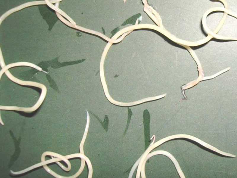 Parasitical Worms.com Illness caused by the nematode of Angiostrongylus cantonensis parasitizes and causes disease in the meninges, invasion of the brain can lead to death. Commonly called Meningitis - brain caused by Angiostrongylus cantonensis. The causative agent of nematode ...
 Fascioliasis is two types of fascioliasis and small liver fluke. People are infected with food, skin. Flukes can cause hepatitis, liver tumors, liver necrosis, but fortunately, liver fluke can be cured if detected early, treated in a reputable facility with a good doctor, using drugs. Good, ...Parasitical Worms.com Diagnosis is determined by seeing sparganum larvae from the wound. Clinical and prehistoric images of frog meat, eye-copying as well as the habit of eating undercooked snakes, mice, and eels are important factors for diagnosis. Doctor: Le Thi Huong Giang Medical Consultation: ...
 MUSHROOM DISEASE (Aspergillus) 1. Epidemiology. Aspergillus fungus is one of the largest fungal strains, present in all over the world, there are about 100 species, currently there are about 20-30 species that cause disease in humans, important strains are A. fumigatus, A. flavus , A. niger such as ...
 MUSHROOM DISEASE Cryptococcosis (Tolurosis, European Blastomycois) 1. Etiology and epidemiology Cryptococcosis is also known as the European Blastomycose mycosis caused by Cryptoccocus neoformans, a thick cystic yeast, has serotypes A, D (C. neoformans var. Neoformans) and B, C ( C.neoformans var. ...MUSHROOM DISEASE Sporotrichosis (Gardener Disease) 1. Epidemiology and etiology Sporotrichosis is a chronic disease caused by Sporothrix schenckii that causes damage to the skin or internal organs (also known as gardener disease - gardener's disease). This is a dimorphic mushroom. In nature, ...CANDIDA MUSHROOM 1. Germs Candidiasis is an acute, subacute or chronic disease caused by Candida-like yeasts, mostly Candida albicans. Candidiasis is available in the body (bronchus, oral cavity, intestine, vagina, skin around the anus) normally in non-pathogenic form. When having favorable ...