Dogiasis Disease, Causes Symptoms And Treatment Of Dogiasis Disease
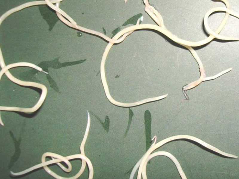
Parasitical Worms.com Adult flukes are very small, 3 - 6 mm long, with 4 suction heads and a double hook, very short neck; coal consists of 3 segments, the final flukes have several hundred eggs, size 45 x 35 mcm, very similar to Toenia spp eggs.
The disease is caused by the larva Echinococcus granulosus, a small intestinal parasite that feeds on meat, mainly dogs.
The causative agent of schistosomiasis
Adult flukes are very small, 3 - 6 mm long, with 4 suction heads and a double hook, very short neck; the body consists of 3 segments, the last flukes have several hundred eggs, size 45 x 35 mcm, very similar to Toenia spp eggs.
Adult dog flukes live in small intestines of dogs, wolves, jackals, mink sometimes cats, and other predators
The eggs, when leaving the host, will be able to live for weeks to months in the outdoors (soil, grass, vegetables, etc
When herbivores swallow a dog fluke, or when a person eats raw vegetables to swallow an egg, the six hookahs get out of the egg and enter the small intestine wall, from which, according to the lymphatic vessels or blood to the heart. and carried it all over the body.
 .
.In places where dog flukes move to (lungs, liver, spleen, brain ...), 6-hook embryos grow to become tumor-shaped larvae (hydatid cyst), after 5 months can be as large as 10mm.
Full-grown growths are usually 1 to 7 cm in diameter, with large bumps up to 20cm
- Laminated membrane: not multiplied, about 1mm thick, including many cuticule layers secreted by the germinal membrane acting as a selective osmosis film.
- The germinal layer, very thin, brittle, made by a layer of syncytium, with nuclei, produces flukes, sporadic wrap.
- Brood capsule (brood capsule), inside the fluke heads.
- Daughter cyst: the germplasm separates from the germinal membrane and secretes the lamina
- Sand fluke (hydatid dand) is the flukes that fall into layers on the bottom of the hump.
 . A fully developed tapeworm can have up to 2,000,000 flukes
- Inside the tumor is a sterile, colorless or pale yellow fluid.
Tapeworm tumors may grow like a benign tumor, often to a degree that growth stops, dies and dries.
In some cases, the tumor loses its head and is no longer contagious (sterile cyst).
Epidemiology of dog flu disease
Dog flukes are common in places where sheep are raised: Alaska, South America, South Africa, Western and Southern Europe, Lebanon, Syria, Algeria, South Australia, New Zealand, Central Asia, and North China Attention should be paid to this parasite when Vietnam started developing sheep-raising sheep, Echinococcus granulosus was found in our country.
People who are closely related to dogs (shepherds) are at a higher risk of infection. Often infected by dog paws, tapeworm eggs stick to their hands and bodies when they eat. Herbivores such as sheep, buffaloes, cows, goats, etc. in fields where dogs are normally seen are highly contaminated.
Clinical manifestations of schistosomiasis
Symptoms of dog tapeworm infection depend on where the parasite is located.
 . It is estimated that:
- 66% of tumors are in liver and peritoneum
- 22% goiter in the neck
- 3% kidney tumors
- 2% of tumors are in bone
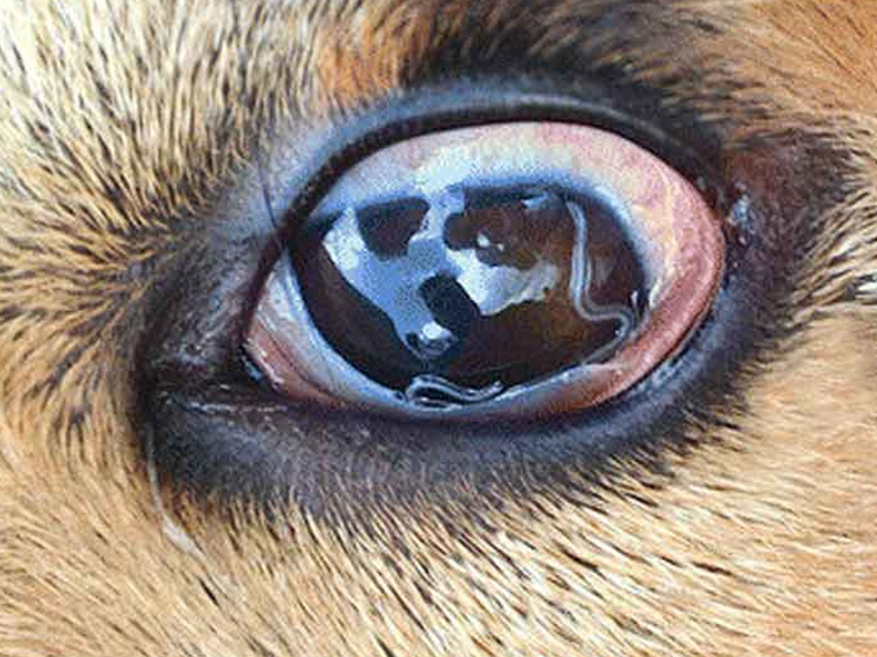
- 6% of tumors are in other organs such as muscles, spleen, eyes, heart, thyroid ..
The host responds by forming fibrous tissue that surrounds the tumor. The tumor grows, squeezing the surrounding tissue, causing the cells to atrophy and die. Blood vessels near the tumor may erode causing bleeding. Tumors in the peritoneum can twisted, constrict blood vessels, leading to necrosis.
More than 75% of liver tumors are found in the right lobe, progressing to the abdomen. Pinched duct can lead to jaundice. When a patient coughs or is physically active, or during surgery, the tumor may burst; flukes are widely spread to other organs, 2 to 5 years after the onset of symptoms of secondary tumors.
When the tumor in the left heart breaks, the fluke heads move to the brain, spleen, kidneys, liver and other organs; The tumor in the heart must allow the fluke to move through the lungs.
The bumps around the trachea often break when the patient coughs, the incomplete rupture and secondary bacterial infection produce chronic apabs.
Brain tumors create syndrome of increased intracranial pressure and Jackson-like epilepsy.
Kidney tumors cause back pain (intermitten pain), blood in the urine, etc.
Tumors in the spleen hurt the ribs and make the ribs rough.
When the hump is located in the pelvis (pelvis), the abdomen will show a murky and echoing zone.
Tumors in the vertebra can cause spinal cord damage.
Tumors in the long bones, pelvis, ribs develop in the gap of bone marrow, gradually eroding bone tissue In the bone there are many small tumors, little or no fluid, nor a fluke. The bones with the parasite become porous, brittle.
 .
.When the tumor bursts, there are allergy symptoms such as itching, hives, then abnormal body temperature, digestive disorders, dyspnea, pale heart, fainting, coma; If fluid drains into the bloodstream, the patient experiences anaphylaxis and may die. Acute leukocyte (BCTT) in the blood increased by about 50% of cases.
After a few years, the tumor died and calcified. Prognosis is good when the tumor is present. . Dịch vụ: Thiết kế website, quảng cáo google, đăng ký website bộ công thương uy tín
Related news
-
 Parasitical Worms.com Tests to find the cause of urticaria, diagnosis of urticaria results will be available throughout the day. After the results the doctor will explain, point out the abnormal signs for your child to understand and he will prescribe medication for home. Question Hello doctor: I ...
 Parasitical Worms.com Adult flukes are very small, 3 - 6 mm long, with 4 suction heads and a double hook, very short neck; coal consists of 3 segments, the final flukes have several hundred eggs, size 45 x 35 mcm, very similar to Toenia spp eggs. The disease is caused by the larva Echinococcus ...
 Parasitical Worms.com Some diseases caused by larvae of the anisakinae family parasitize marine mammals. In humans, the parasite falls into a dead-end, or severe or severe illness depending on the place of parasite, number of larvae and tissue responses. Diagnosis is often difficult and the most ...
 Parasitical Worms.com Illness caused by the nematode of Angiostrongylus cantonensis parasitizes and causes disease in the meninges, invasion of the brain can lead to death. Commonly called Meningitis - brain caused by Angiostrongylus cantonensis. The causative agent of nematode ...
 Fascioliasis is two types of fascioliasis and small liver fluke. People are infected with food, skin. Flukes can cause hepatitis, liver tumors, liver necrosis, but fortunately, liver fluke can be cured if detected early, treated in a reputable facility with a good doctor, using drugs. Good, ...
 Parasitical Worms.com Diagnosis is determined by seeing sparganum larvae from the wound. Clinical and prehistoric images of frog meat, eye-copying as well as the habit of eating undercooked snakes, mice, and eels are important factors for diagnosis. Doctor: Le Thi Huong Giang Medical Consultation: ...
 MUSHROOM DISEASE (Aspergillus) 1. Epidemiology. Aspergillus fungus is one of the largest fungal strains, present in all over the world, there are about 100 species, currently there are about 20-30 species that cause disease in humans, important strains are A. fumigatus, A. flavus , A. niger such as ...
 MUSHROOM DISEASE Cryptococcosis (Tolurosis, European Blastomycois) 1. Etiology and epidemiology Cryptococcosis is also known as the European Blastomycose mycosis caused by Cryptoccocus neoformans, a thick cystic yeast, has serotypes A, D (C. neoformans var. Neoformans) and B, C ( C.neoformans var. ...
 MUSHROOM DISEASE Sporotrichosis (Gardener Disease) 1. Epidemiology and etiology Sporotrichosis is a chronic disease caused by Sporothrix schenckii that causes damage to the skin or internal organs (also known as gardener disease - gardener's disease). This is a dimorphic mushroom. In nature, ...
 CANDIDA MUSHROOM 1. Germs Candidiasis is an acute, subacute or chronic disease caused by Candida-like yeasts, mostly Candida albicans. Candidiasis is available in the body (bronchus, oral cavity, intestine, vagina, skin around the anus) normally in non-pathogenic form. When having favorable ...


