Screening And Treatment Of Neurophotic Disease In Hcmc
Parasitical Worms.com Pinworm was first described by Linnaeus in 1758 and was the sole host. In 1983, Hugot isolated another pinworm living on the human, Enterobius gregorii. However, some authors argue that this is only a development stage of E.vermicularis
With a specific development cycle, pinworms have expressed their specific features in terms of epidemiology, pathology as well as disease control, different from other worms of the intestinal parasite.
The development cycle of pinworms
1. THE FIGURE OF KIMUN
Adult pinworms
Pinworms are ivory-white, cylindrical, the head has three lips surrounding the mouth The end of the esophagus is slightly dilated before enlarging into a round, bulbous mound, adjacent to the intestine.
The cuticle on both sides of the body thickens at the head to form two wings (alae), then gradually narrows into two ridges running along the length of the body and is the characteristic to identify adult pinworms on the tissue samples.
 . Cross sectional surgery. The size of the female worm is about 8 - 13mm x 0.3 - 0.5mm, with a pointed and straight tail, accounting for nearly one third of the body length. Smaller male worms (2-5mm x 0
Pinworm eggs
Pinworm eggs are oval, unilateral, about 50 - 60 xm x 20 - 30 µm, transparent shell, relatively thick, consisting of sticky outer layer of albumine, followed by two layers of chitine and a lipid film innermost.
The outer layer has a mechanical protective function while the role of the lipid layer is to resist chemical agents.
2. DEVELOPMENT PROCESS OF KIM GIUN
Adult worms parasitize mainly in the cecum.
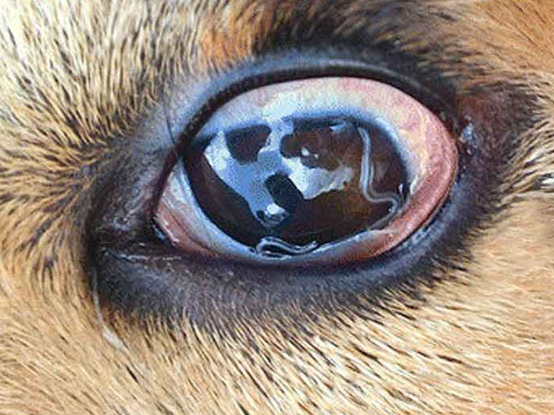 . However, in severe infections, they will invade adjacent parts such as the ileum, colon. Worms can roam freely in the stomach of the stomach to the anus or attach to the lining of the intestine with three lips.
Pinworm life is very short, only about 4-13 weeks for females, 2-7 weeks for males.
After intercourse, the male worms die immediately, the female worm moves gradually to the anus with a uterus filled with embryonic eggs Eggs require oxygen to fully develop to the stage of infection.
So at night, the female worms swim to the edge of the anus to lay eggs and die after giving birth. The average number of eggs released from a female worm is 11,000 eggs, which can vary from 4,000 to 17,000.
The albumin shell helps the egg to attach to the skin around the anus. Under the right temperature conditions (35-420C), after 4 - 8 hours, the embryo eggs will develop into eggs containing infected larvae.
Below 220C and above 460C eggs do not develop.
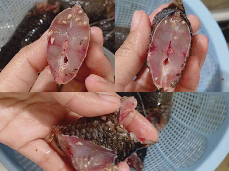 . Eggs from the edge of the anus or sticking on the hands of an infected person will spread around the patient's living space (beds, mats, blankets, floors, pajamas, toys, doorknobs, etc.)
In addition, pinworm eggs are very light, can follow the diffuse dust into the air in the room. With high humidity and room temperature, an infected egg can survive for 2-3 weeks outside the human body, but only for a few hours or days in dry conditions.
Pollution of eggs in the environment around the host has created favorable conditions for pathogens to infect other members living with patients.
When a person swallows an infected egg, the larvae are released in the duodenum, molt twice into young worms, and then move down to the cecum to mature. The time from swallowing eggs containing larvae to adult worms laying eggs takes about 4 - 8 weeks.
The characteristic that eggs are laid at the edge of the anus and quickly become infectious leads to reinfection during the development of pinworms:
The habit of holding hands is to create opportunities to swallow the eggs stuck on the hands of patients after scratching the anus; Although rare, the larvae may hatch around the anus and then crawl up to the rectum, causing retroinfection.
3. Characteristics of pinworm disease
Characteristics in the development cycle show that the distribution of pinworm disease depends mainly on the issue of personal hygiene rather than geography and climate.
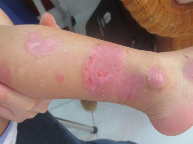 Therefore, the disease is widespread, especially in crowded areas, cramped living conditions and poor sanitation.
How do you and your child get pinworm infection and anal itching?
Female pinworms containing embryos around the anus spread to the indoor environment, you and your child will swallow eggs through their hands, toy food ... infected pinworm eggs, can also breathe dust containing eggs, The egg then enters the digestive tract and parasites in the large intestine.
However, in general, the prevalence of disease predominates in temperate countries rather than tropical countries. Perhaps, in cold countries, people are less likely to change clothes and not to shower frequently to facilitate disease development and spread.
On the other hand, in tropical countries Pinworm infection is less of a concern for research than other important helminths.
Worldwide, an estimated 200 million people are infected with vermicularic Enterobius each year.
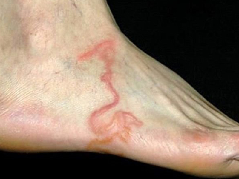 . The prevalence rate in some Asian countries varies from 21% - 74%. In Vietnam, the disease ranges from 7% - 50%, in some places up to 80% - 100%. Surveys in Cu Chi district, Ho Chi Minh City in 2007-2009 recorded about 22.7% - 53.5% of preschoolers infected with helminths. . Dịch vụ: Thiết kế website, quảng cáo google, đăng ký website bộ công thương uy tín
Related news
-
 Parasitical Worms.com Tests to find the cause of urticaria, diagnosis of urticaria results will be available throughout the day. After the results the doctor will explain, point out the abnormal signs for your child to understand and he will prescribe medication for home. Question Hello doctor: I ...
 Parasitical Worms.com Adult flukes are very small, 3 - 6 mm long, with 4 suction heads and a double hook, very short neck; coal consists of 3 segments, the final flukes have several hundred eggs, size 45 x 35 mcm, very similar to Toenia spp eggs. The disease is caused by the larva Echinococcus ...
 Parasitical Worms.com Some diseases caused by larvae of the anisakinae family parasitize marine mammals. In humans, the parasite falls into a dead-end, or severe or severe illness depending on the place of parasite, number of larvae and tissue responses. Diagnosis is often difficult and the most ...
 Parasitical Worms.com Illness caused by the nematode of Angiostrongylus cantonensis parasitizes and causes disease in the meninges, invasion of the brain can lead to death. Commonly called Meningitis - brain caused by Angiostrongylus cantonensis. The causative agent of nematode ...
 Fascioliasis is two types of fascioliasis and small liver fluke. People are infected with food, skin. Flukes can cause hepatitis, liver tumors, liver necrosis, but fortunately, liver fluke can be cured if detected early, treated in a reputable facility with a good doctor, using drugs. Good, ...
 Parasitical Worms.com Diagnosis is determined by seeing sparganum larvae from the wound. Clinical and prehistoric images of frog meat, eye-copying as well as the habit of eating undercooked snakes, mice, and eels are important factors for diagnosis. Doctor: Le Thi Huong Giang Medical Consultation: ...
 MUSHROOM DISEASE (Aspergillus) 1. Epidemiology. Aspergillus fungus is one of the largest fungal strains, present in all over the world, there are about 100 species, currently there are about 20-30 species that cause disease in humans, important strains are A. fumigatus, A. flavus , A. niger such as ...
 MUSHROOM DISEASE Cryptococcosis (Tolurosis, European Blastomycois) 1. Etiology and epidemiology Cryptococcosis is also known as the European Blastomycose mycosis caused by Cryptoccocus neoformans, a thick cystic yeast, has serotypes A, D (C. neoformans var. Neoformans) and B, C ( C.neoformans var. ...
 MUSHROOM DISEASE Sporotrichosis (Gardener Disease) 1. Epidemiology and etiology Sporotrichosis is a chronic disease caused by Sporothrix schenckii that causes damage to the skin or internal organs (also known as gardener disease - gardener's disease). This is a dimorphic mushroom. In nature, ...
 CANDIDA MUSHROOM 1. Germs Candidiasis is an acute, subacute or chronic disease caused by Candida-like yeasts, mostly Candida albicans. Candidiasis is available in the body (bronchus, oral cavity, intestine, vagina, skin around the anus) normally in non-pathogenic form. When having favorable ...

