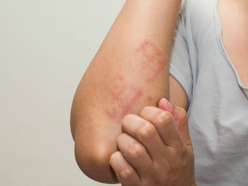
Is Amip Hazardous?
IS AMIP DISEASE RISK NO
Entamoeba histolytica is popular all over the world, especially in tropical countries, hot and humid climate, Entamoeba histolytica parasitic in the colon, amoeba in humans capable of causing true disease. Contagious disease caused by ingestion of follicles present in food and drinking water. Most cases have no symptoms, if any, common symptoms are amoebic dysentery. In addition, amoeba can cause disease in other organs such as liver abscess, lung abscess - pleura .
Amoeba causes liver abscess
1. AMIP DISEASES
Entamoeba histolytica consists of three forms:
Histolycia form
Also known as an active erythrocyte size 20µm - 40µm, moving thanks to the artificial leg in a certain direction
 .5 µm - 9.0 µm. The active body has a single circular nucleus, diameter 4 --m - 7 µm; Chromatin particles are regularly arranged in the nuclear membrane; The nucleus has a nucleus in the middle of the diameter of 0.5 µm.
Amoeba causes lung abscess
It is a pathogenic, invasive tissue, thanks to the enzyme that destroys proteins and reproduces by dividing
The minuta
Also known as non-erythrocyte active form size 10 - 12 ,m, smaller than histolytica, living saprophytic in the large intestine due to food residues, germs. The minuta form can turn into cystic form or be able to eat red blood cells depending on living conditions in the large intestine.
Cystic form.
Spherical or slightly oval, size 10-16µm, refractive cyst wall and relatively thick 125 - 150nm so that the cystic body is resistant to the outside.
 . Cytoplasm has 1 to 4 nuclei, the nucleus is in the middle of the nucleus; vacuoles contain glycogen, this vacuole tends to disappear when the follicle matures (quadrilateral cyst). In addition, it can be buried (chrimatoid form) due to ribosome gathered, stick-shaped or round ends.
This form finds a teacher in the stool and is able to spread disease from person to person.
Shape of amoeba
2 AMIP DISEASE DEVELOPMENT PROCESS
Consists of two cycles: no pathogenesis (in the intestine) and pathogenesis (large intestine wall and tissue).
The cycle does not cause disease.
People are infected with Entamoeba histolytica by ingesting four-cell cysts through the digestive tract. Upon reaching the stomach, the cyst is not altered by the gastric acid environment, so there is never an active infection through the gastrointestinal tract. On the other hand, the active body dies quickly when it comes out of the body for more than 2 hours. When the cyst reaches the small intestine, the neutral or slightly alkaline pH destroys the cyst wall and divides the nucleus.
 . The cytoplasm then divides, creating eight minuta bodies.
When the environment in the large intestine becomes unfavorable, for example, dehydration .. the minuta can shrink and become immobile, forming a cystic precursor. The follicle makes its own wall and becomes a follicle. The follicles follow the stool. When externally located, the single-follicular cyst continues to develop into a double-nucleus follicle and then becomes the progenitor cell. The four-core cyst itself is the stage of infection.
In the case of the large intestine, the stool becomes concentrated, dehydrated, at which time a single-follicular cyst also develops rapidly into two, four cores and then follows the stool out.

When there are many advantages, such as decreased host resistance, infection ... the minuta body will turn into a pathogenic form - able to eat red blood cells.
Pathogenesis cycle.
The active form of eating erythrocytes has many enzymes (trypsine, pepsine, hyaluronidase ..) causing activity, hemorrhage ..
.., they invade the large intestine wall, reproduce by dividing in half. From the intestinal wall, amoeba can travel through the bloodstream to other organs (liver, lungs, brain ...) and continue to reproduce to cause diseaseThe cycle of development of amoeba
3. EPIDEMIOLOGICAL CHARACTERISTICS AMIP
Amoeba infection is common everywhere, especially in tropical countries.
People are infected with amoeba due to ingestion of cysts in food and drinking water.
Thanks to the thick wall, the cysts are resistant to entry.
. Cysts can survive in soil for 8 days at 28-34 ° C, 40 days at 2-6 ° C. Cysts are destroyed when boiled or exposed to acetic acid 5% - 10%.People who are the owners of E. histolytica, especially those who are amoeba-carriers, have no clinical manifestations
Cysts can live long in the external environment, so the external environment is also the host of this parasite.
The main sources of infection are:
Water is contaminated with human feces.
Fresh vegetables have infected human feces.
Flies and cockroaches help transport germs from feces to food.
Currently, the disease is also transmitted through sexual contact in gay men.
4.
 . Clinical manifestations of AMIP
Amoeba intestinal.
Amoebic dysentery
Clinical
Relatively long incubation period (1-4 weeks), the patient may have diarrhea and abdominal pain, then the syndrome appears:
Severe abdominal pain, pain along the colon frame due to amoeba parasite in the colon site usually in the cecum.
The stool is viscous and bloody (<10 times / day) because the amoeba enters the intestinal membrane. Normally there are fish in intestinal membrane. . Dịch vụ: Thiết kế website, quảng cáo google, đăng ký website bộ công thương uy tín
Related news
-
 Parasitical Worms.com Tests to find the cause of urticaria, diagnosis of urticaria results will be available throughout the day. After the results the doctor will explain, point out the abnormal signs for your child to understand and he will prescribe medication for home. Question Hello doctor: I ...Parasitical Worms.com Adult flukes are very small, 3 - 6 mm long, with 4 suction heads and a double hook, very short neck; coal consists of 3 segments, the final flukes have several hundred eggs, size 45 x 35 mcm, very similar to Toenia spp eggs. The disease is caused by the larva Echinococcus ...
 Parasitical Worms.com Some diseases caused by larvae of the anisakinae family parasitize marine mammals. In humans, the parasite falls into a dead-end, or severe or severe illness depending on the place of parasite, number of larvae and tissue responses. Diagnosis is often difficult and the most ...Parasitical Worms.com Illness caused by the nematode of Angiostrongylus cantonensis parasitizes and causes disease in the meninges, invasion of the brain can lead to death. Commonly called Meningitis - brain caused by Angiostrongylus cantonensis. The causative agent of nematode ...Fascioliasis is two types of fascioliasis and small liver fluke. People are infected with food, skin. Flukes can cause hepatitis, liver tumors, liver necrosis, but fortunately, liver fluke can be cured if detected early, treated in a reputable facility with a good doctor, using drugs. Good, ...
 Parasitical Worms.com Diagnosis is determined by seeing sparganum larvae from the wound. Clinical and prehistoric images of frog meat, eye-copying as well as the habit of eating undercooked snakes, mice, and eels are important factors for diagnosis. Doctor: Le Thi Huong Giang Medical Consultation: ...
 MUSHROOM DISEASE (Aspergillus) 1. Epidemiology. Aspergillus fungus is one of the largest fungal strains, present in all over the world, there are about 100 species, currently there are about 20-30 species that cause disease in humans, important strains are A. fumigatus, A. flavus , A. niger such as ...MUSHROOM DISEASE Cryptococcosis (Tolurosis, European Blastomycois) 1. Etiology and epidemiology Cryptococcosis is also known as the European Blastomycose mycosis caused by Cryptoccocus neoformans, a thick cystic yeast, has serotypes A, D (C. neoformans var. Neoformans) and B, C ( C.neoformans var. ...
 MUSHROOM DISEASE Sporotrichosis (Gardener Disease) 1. Epidemiology and etiology Sporotrichosis is a chronic disease caused by Sporothrix schenckii that causes damage to the skin or internal organs (also known as gardener disease - gardener's disease). This is a dimorphic mushroom. In nature, ...CANDIDA MUSHROOM 1. Germs Candidiasis is an acute, subacute or chronic disease caused by Candida-like yeasts, mostly Candida albicans. Candidiasis is available in the body (bronchus, oral cavity, intestine, vagina, skin around the anus) normally in non-pathogenic form. When having favorable ...