Fascioliasis: Laboratory Test For Trichotomiasis
General information about liver fluke disease
What is a liver fluke?
Flukes, which are infected by humans, include two types, the great fluke and the small fluke. They enter the human body through the gastrointestinal tract, causing pathology in many organs such as the liver, bile ducts and bile ducts. Liver fluke usually causes liver tumors and can lead to liver cancer if left untreated.
The procedure of testing liver fluke at the parasite clinic
After a physical examination, the doctor will order diagnostic tests for liver fluke. In the early stages of infection, the liver fluke eggs do not appear in the stool, so it is not possible to conduct stool tests to diagnose liver fluke at this stage
A blood test using ELISA is the best method to diagnose liver fluke at an early stage of the disease. Blood samples were taken intravenously about 2ml. Then centrifuge plasma and perform on automatic immune testing machine
Subclinical methods are performed when needed
Abdominal ultrasound: images of honeycomb parenchymal lesions or subarctic liver fluid, abscess images Liver flukes on ultrasound can be observed with a leaf-like, flat image if large in size. Abdominal ultrasound together with a stool examination is selected as a screening test for disease in areas with high levels of liver fluke infection.
 .
.Computerized tomography of the abdomen: survey of bile ducts better than abdominal ultrasound. Expansion of the bile ducts, without obstruction, is a typical image in liver fluke.
Fascioliasis is a long-term infection with liver necrosis
The procedure to treat liver fluke in the parasite clinic
After the test results are available, you will see your doctor to analyze the results and prescribe an infection.
Treatment of liver fluke not liver and biliary tract damage
The early stage of liver fluke in the digestive tract, has not moved to the bile, so the treatment time for liver fluke is shorter, easier to treat. Only use flukes kill drugs without antibiotics
Note that some drugs that treat liver fluke are less effective or drug-resistant liver fluke, the treating physician should have experience in order to use the best and most effective medicine for tapeworm infection at this stage Early treatment prevents them from moving to the liver and bile.
Treating liver fluke disease causing complications of hepatitis and biliary tract
At this stage, the liver fluke has moved to the liver and enters the bile ducts in the liver, often causing liver tumors and purulent hepatitis. Therefore the doctor will use antibiotics combined with drugs to treat liver fluke to kill fluke to treat liver fluke at this stage.
If not treated, can I cure liver fluke?
The liver fluke lives in the body, mainly in the liver and in the bile ducts.
. Fascioliasis is a chronic disease that can last for decades, including infection with large fluke and small liver fluke.Fascioliasis is detected in many countries around the world
People infected with small fluke are found in many parts of the world. The species of Opisthorchis viverrini causes about 3 million people in Laos, Cambodia, Thailand and the South of Vietnam. Meanwhile, Clonorchis sinensis is a small liver fluke distributed mainly in Japan, China, Taiwan and the northern provinces of our country
Schistosomiasis spreads from north to south in Vietnam. It is reported that there are about 21 provinces and cities, the infection rates vary by region, the highest in Binh Dinh, Phu Yen, Nam Dinh, Ninh Binh.
Infection with Fasciola hepatica is common in Europe, South America and Africa; while Fasciola gigantica is mainly distributed in Asia. In our country, Fascioliasis is more common than small liver fluke with more than 40 provinces and cities, the highest in the Central region and Central Highlands.
Blue color are common areas of liver fluke infection
What causes liver fluke disease?
Flukes are divided into two groups: small liver fluke and small liver fluke. Small liver fluke has 3 types including: Clonorchis sinensis; Opisthorchis viverrini; Opisthorchis felineus.
 . Fascioliasis has 2 types, including: Fasciola hepatica; Fasciola gigantica.
Characteristics of large liver flukes:
This is a parasite shaped like a leaf, flat body should be called liver fluke Fascioliasis is much larger in size than small liver fluke, about 30 x 10 - 12mm. Liver flukes with male and female genitalia on one body should be classified as bisexual.
Flukes are released into the environment with a thin shell, so they are easily damaged. Water environment is required for eggs to develop into larvae and adult tapeworms to cause disease. In terrestrial environments, under the influence of sunlight, both eggs and adult flukes are very easy to die, hard to survive.
Flukes are major infections for herbivores such as sheep and cattle. Human liver fluke infection is just a coincidence because the person eats raw aquatic plants such as watercress, celery, cilantro or drinking contaminated water.
The main hosts of small liver fluke include human, dog, cat, otter, invasive process and disease.
 . Dịch vụ: Thiết kế website, quảng cáo google, đăng ký website bộ công thương uy tín
Related news
-
 Parasitical Worms.com Tests to find the cause of urticaria, diagnosis of urticaria results will be available throughout the day. After the results the doctor will explain, point out the abnormal signs for your child to understand and he will prescribe medication for home. Question Hello doctor: I ...Parasitical Worms.com Adult flukes are very small, 3 - 6 mm long, with 4 suction heads and a double hook, very short neck; coal consists of 3 segments, the final flukes have several hundred eggs, size 45 x 35 mcm, very similar to Toenia spp eggs. The disease is caused by the larva Echinococcus ...
 Parasitical Worms.com Some diseases caused by larvae of the anisakinae family parasitize marine mammals. In humans, the parasite falls into a dead-end, or severe or severe illness depending on the place of parasite, number of larvae and tissue responses. Diagnosis is often difficult and the most ...
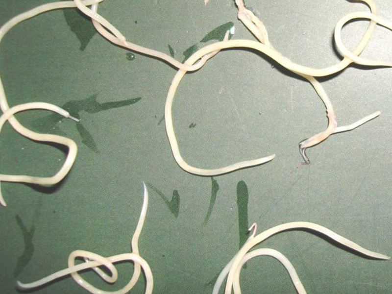 Parasitical Worms.com Illness caused by the nematode of Angiostrongylus cantonensis parasitizes and causes disease in the meninges, invasion of the brain can lead to death. Commonly called Meningitis - brain caused by Angiostrongylus cantonensis. The causative agent of nematode ...
 Fascioliasis is two types of fascioliasis and small liver fluke. People are infected with food, skin. Flukes can cause hepatitis, liver tumors, liver necrosis, but fortunately, liver fluke can be cured if detected early, treated in a reputable facility with a good doctor, using drugs. Good, ...Parasitical Worms.com Diagnosis is determined by seeing sparganum larvae from the wound. Clinical and prehistoric images of frog meat, eye-copying as well as the habit of eating undercooked snakes, mice, and eels are important factors for diagnosis. Doctor: Le Thi Huong Giang Medical Consultation: ...
 MUSHROOM DISEASE (Aspergillus) 1. Epidemiology. Aspergillus fungus is one of the largest fungal strains, present in all over the world, there are about 100 species, currently there are about 20-30 species that cause disease in humans, important strains are A. fumigatus, A. flavus , A. niger such as ...
 MUSHROOM DISEASE Cryptococcosis (Tolurosis, European Blastomycois) 1. Etiology and epidemiology Cryptococcosis is also known as the European Blastomycose mycosis caused by Cryptoccocus neoformans, a thick cystic yeast, has serotypes A, D (C. neoformans var. Neoformans) and B, C ( C.neoformans var. ...
 MUSHROOM DISEASE Sporotrichosis (Gardener Disease) 1. Epidemiology and etiology Sporotrichosis is a chronic disease caused by Sporothrix schenckii that causes damage to the skin or internal organs (also known as gardener disease - gardener's disease). This is a dimorphic mushroom. In nature, ...CANDIDA MUSHROOM 1. Germs Candidiasis is an acute, subacute or chronic disease caused by Candida-like yeasts, mostly Candida albicans. Candidiasis is available in the body (bronchus, oral cavity, intestine, vagina, skin around the anus) normally in non-pathogenic form. When having favorable ...