Is Small Laryngeal Disease Dangerous?
There are 3 species of small liver fluke: Clonorchis sinensis, Opisthorchis viverrini and Opisthorchis felineus. The distribution of these small liver fluke species is also very different; small liver fluke Clonorchis sinensis (C.sinensis).
The disease is common in China, Korea, Hong Kong, Japan, Russia and some Southeast Asian countries such as the Philippines, Singapore, Malaysia and Northern Vietnam.
The small liver fluke Opisthorchis viverrini (O
- For Vietnam since 1908 Mouzel, 1909 Mathis and Leger have found Csinensis. In 1924 Railiet discovered O.
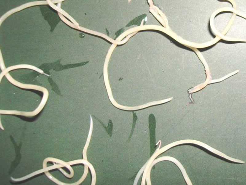 .felineus in Hanoi. In 1965 Dang Van Ngu and Do Duong Thai encountered an case of O.felineus in collaboration with C. Sinin.
A- Opisthorchis viverrini
B- Opisthorchis felineus
C- Clonorchis sinensis
1
11. Flukes mature.
C.sinensis small liver fluke has leaf shape, flat body, light red color.
 . Flukes 10-12mm long, 2-4mm wide, with two suction mouths, anterior suction mouth connected to the gastrointestinal tract with a diameter of 600 µm, rear suction mouth (mouth sucking mouth) with a diameter of 500µm. The digestive tract runs along the sides of the body and is a blocked tube, not connected together. Flukes do not have anus because the fluke's nutrition mainly penetrates the nutrients through its surface. Therefore, there are many nutritional glands on the flukes
On the flukes there are both male and female genitalia. Testicular branching, almost all behind the body. The ovary is about the middle of the body, the uterus runs zigzag, contains many eggs.
The picture of a small liver fluke parasite in the liver
1.2. Egg
The liver fluke eggs are very small, size 16-17µm x 26-30µm, oval, one pole with a cap shaped like a cap, a bigger bulge like a balloon with a small bottom and spines.
 . Dark yellow eggs. The skin is thin, smooth and has a double border Inside the nucleus may have larval images.
2. Development cycle
2.1. Parasitic position
Flukes of the liver are parasitic in the small bile ducts in the liver. If abundant, the fluke can destroy liver parenchyma and parasitize the liver. Flukes by absorbing nutrients from bile.
2.
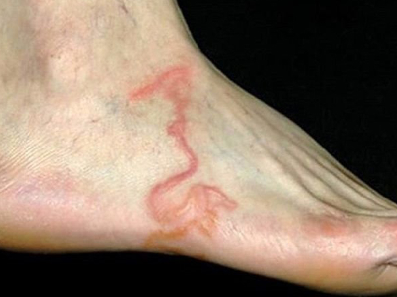 2. Evolution of the cycle
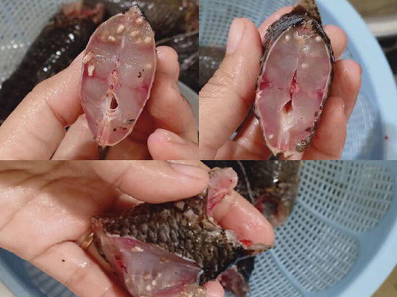
Flukes of the liver lay eggs in the bile ducts. Eggs follow the bile to the intestine and follow the stools. Eggs need a water environment to develop into hairy larvae. The water-free swimming larvae find parasites in some snails to develop into larvae (tail larvae) 21 to 30 days after entering the snails. Hairy larvae live in the intestine, liver, pancreas of snails. Types of snails that are suitable hosts for larvae include snails of the genus Bythinia, Bythinia snails have many types, mainly in China, Japan, Korea, Taiwan. After that, the larvae leave the snails to freshwater fish to develop into larval follicles in the flesh of the fish, this is the stage of infection In Vietnam, common carp, perch, crucian, drift fish ..
 .. can all be intermediate hosts of small liver fluke.
When humans or animals (cats, dogs ..) eat raw fish (raw fish salad) or undercooked fish, the larvae will follow the food into the intestinal tract after 15 hours, move to the bile duct to the liver and after 26 days will develop. adult flukes and pathogens
Small liver fluke Clonorchis sinensis can live 15-25 years in the human body.
3. Pathology
3.1.
 . Pathological lesions
Flukes of the liver cause serious damage to the liver. Flukes cause frequent irritation to the liver, while occupying food and toxic. The parasitic position and size of the flukes are prone to occlusion.
Because the flukes usually adhere to the bile duct, use their mouths to suck food, the liver will gradually spread sclerosis at the door, the liver organ will be proliferated and may lead to liver fibrosis, ascites, regression Fats in the liver
Toxins secreted by tapeworm can cause allergic conditions, sometimes can cause anemia, eosinophilia.
Regarding pathological lesions, the liver is markedly enlarged. The weight of the liver can be up to 4kg. On the liver, there are bulging spots, the bulges are usually pale white and correspond to the expansion of the bile duct. If you cut the bulging points, you will see a gray-green fluid.
3.
 .2. Symptom
Clinical manifestations of small liver fluke depend on the intensity of infection and the response of the host In rare cases, there are no special symptoms. In case of liver fluke infection with more than 100 children, the disease manifests itself through 2 stages:
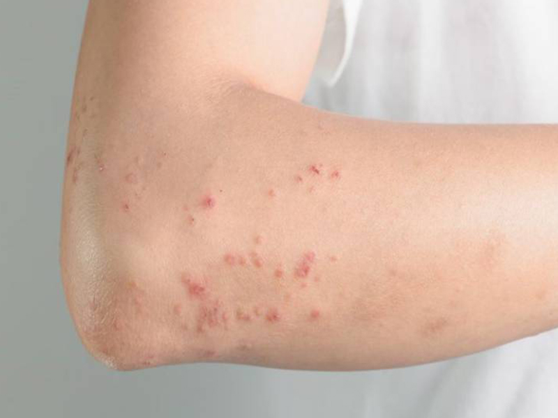
The onset of illness usually begins with signs of gastrointestinal disorders such as anorexia, indigestion, dull pain in the liver, diarrhea or constipation. Attached can see the whole rash, rash.
The full-onset stage involves more pain in the liver, accompanied by anemia, jaundice and ascites, which may occur later. If there is a bacterial superinfection, the patient may have fever in episodes or a prolonged fever.
4. Diagnosis
4.1.
 Clinical diagnosis
Nonspecific because it is easily confused with other biliary diseases.
4.2. Diagnostic tests
- Stool test for eggs by direct technique or egg collection. A stool test for flukes is a simple but confirmed method of getting the disease,
- In case of infection. . Dịch vụ: Thiết kế website, quảng cáo google, đăng ký website bộ công thương uy tín
Related news
-
 Parasitical Worms.com Tests to find the cause of urticaria, diagnosis of urticaria results will be available throughout the day. After the results the doctor will explain, point out the abnormal signs for your child to understand and he will prescribe medication for home. Question Hello doctor: I ...
 Parasitical Worms.com Adult flukes are very small, 3 - 6 mm long, with 4 suction heads and a double hook, very short neck; coal consists of 3 segments, the final flukes have several hundred eggs, size 45 x 35 mcm, very similar to Toenia spp eggs. The disease is caused by the larva Echinococcus ...
 Parasitical Worms.com Some diseases caused by larvae of the anisakinae family parasitize marine mammals. In humans, the parasite falls into a dead-end, or severe or severe illness depending on the place of parasite, number of larvae and tissue responses. Diagnosis is often difficult and the most ...
 Parasitical Worms.com Illness caused by the nematode of Angiostrongylus cantonensis parasitizes and causes disease in the meninges, invasion of the brain can lead to death. Commonly called Meningitis - brain caused by Angiostrongylus cantonensis. The causative agent of nematode ...
 Fascioliasis is two types of fascioliasis and small liver fluke. People are infected with food, skin. Flukes can cause hepatitis, liver tumors, liver necrosis, but fortunately, liver fluke can be cured if detected early, treated in a reputable facility with a good doctor, using drugs. Good, ...
 Parasitical Worms.com Diagnosis is determined by seeing sparganum larvae from the wound. Clinical and prehistoric images of frog meat, eye-copying as well as the habit of eating undercooked snakes, mice, and eels are important factors for diagnosis. Doctor: Le Thi Huong Giang Medical Consultation: ...
 MUSHROOM DISEASE (Aspergillus) 1. Epidemiology. Aspergillus fungus is one of the largest fungal strains, present in all over the world, there are about 100 species, currently there are about 20-30 species that cause disease in humans, important strains are A. fumigatus, A. flavus , A. niger such as ...
 MUSHROOM DISEASE Cryptococcosis (Tolurosis, European Blastomycois) 1. Etiology and epidemiology Cryptococcosis is also known as the European Blastomycose mycosis caused by Cryptoccocus neoformans, a thick cystic yeast, has serotypes A, D (C. neoformans var. Neoformans) and B, C ( C.neoformans var. ...
 MUSHROOM DISEASE Sporotrichosis (Gardener Disease) 1. Epidemiology and etiology Sporotrichosis is a chronic disease caused by Sporothrix schenckii that causes damage to the skin or internal organs (also known as gardener disease - gardener's disease). This is a dimorphic mushroom. In nature, ...
 CANDIDA MUSHROOM 1. Germs Candidiasis is an acute, subacute or chronic disease caused by Candida-like yeasts, mostly Candida albicans. Candidiasis is available in the body (bronchus, oral cavity, intestine, vagina, skin around the anus) normally in non-pathogenic form. When having favorable ...


