Parasitic Sarcocystis Spp Causes Food Poison
Parasitic SARCOCYSTIS SPP CAUSES FOOD POISON
1. SARCOCYSTIS PARAMETERIC SIGNATURE
Sarcocystis hominis is a protozoan parasite that infects humans through the gastrointestinal tract and causes illness in humans with manifestations in dangerous muscles, bones, and organs.
In the fresh feces, the spore-like form of the follicle is irregularly discharged, in small numbers, the oval is long, symmetrical on both ends, the size 14-15 15m x 9-10 µm, colorless, transparent, thick outer shell and very refractive, inside contains four sickle-shaped applicators, converging to one end of the follicle spores, whose nucleus lies in the first 1/3 - 1/3 middle, while the awl is at the opposite end, creating into separate large particles. Sometimes a large number of follicular spores are released in liquid faeces.
Nang Sarcocystis hominis in muscle
Sarcocystis cysts in the body are common in skeletal muscle, tongue muscle, heart muscle, sometimes in the larynx, pharynx, upper esophagus, spherical capsule, thick wall, contain disinfection, need to distinguish with the toxoplasmosis gondii capsule using molecular biology tests
2. SARCOCYSTIS PARENTAL DEVELOPMENT PROCESS
The complete cycle occurs on two different hosts
- Cycle of paralysis occurs on intermediate hosts (KCTG) as herbivores.
- The gametocyte cycle occurs on the permanent host (KCVV) as predators, this cycle takes place in the small intestinal epithelial cells, creating follicles and follicles according to the stool KCVV to the outside , developed to produce two follicular spores, each of which has four topical application
When KCTG swallows the follicle spores, the parasite is released, invading the small intestinal mucosa, into the endothelial cells, reproducing asexually, creating many pieces of germ These fragments can invade skeletal muscle and the heart muscle, creating cysts. Follicular cysts in the muscles are reservoirs that can infect parasites.
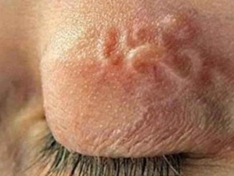 .
.People are infected by eating undercooked pork or beef, the intestines infiltrate the small intestinal epithelial cells, transforming into male and female gametes.
After fertilization, the zygote is transformed into a mature follicle into an adult follicle containing two follicles, each containing four germs. The follicle ruptures in the intestine and the follicle spreads in the feces outward. Double
when humans are the KCTG of Sarcocystis of other animals.
3
The disease is caused by muscle sarcocystis
Studies show a high incidence of overall disease in Southeast Asia, Wong et al 1992, autopsy of tongue muscles in humans, a positive rate of about 21% between 16 and 59 years of age. . In addition to tongue muscles, the muscles in the head and neck are also common. Nearly 100 cases were recorded in the literature.
 .
.There is evidence that the person acting as the intermediate host of the Sarcocystis species should have been infected only in monkeys. In 1993, a US army involved in a civilian project in Malaysia suffered from an outbreak of eosinophilia caused by Sarcocystis (Arness et al., 1999).
The reason these people are exposed to mud It is also caused by drinking unboiled water, eating indigenous foods, including raw vegetables contaminated with faeces, in the fecal spore of Sarcocystis.
Diseases caused by intestinal sarcocystis
A study by Pena and colleagues in 2001 showed that 100% of Kibbe meat samples living in Sao Paulo restaurant were contaminated with Sarcocystis. Frequent co-infection between Sarcocystis hominis and Sarcocystis cruzi.
Kibbe is a traditional Lebanese dish, thinly sliced beef or lamb seasoning, eaten raw. In volunteers, eating raw Kibbe, then having symptoms of digestive disorders.
Stool test for Thai workers, especially those in the Northern provinces, has a 23% infection rate.
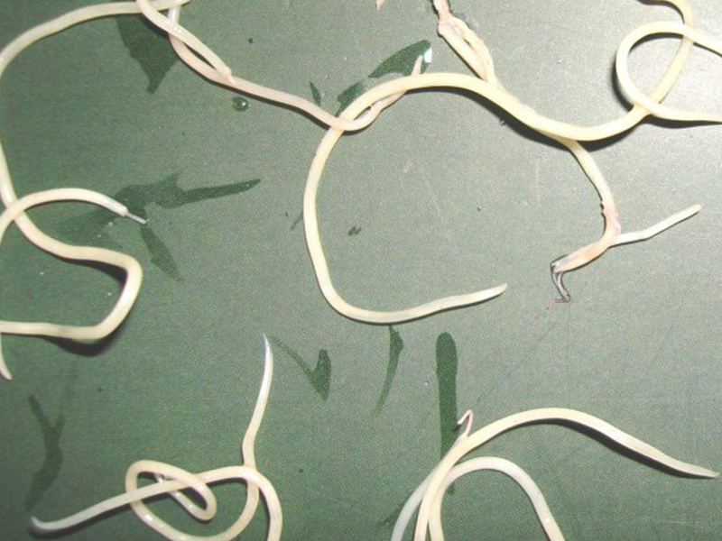 . High prevalence among workers who eat raw meat, have low living standards and poor hygiene; Military people and tourists are also highly vulnerable.
4 Clinical manifestations of SARCOCYSTIS
Most Sarcocystis infections are asymptomatic, but there are also cases of severe illness in patients with poor resistance.
Sarcocystis disease in muscles
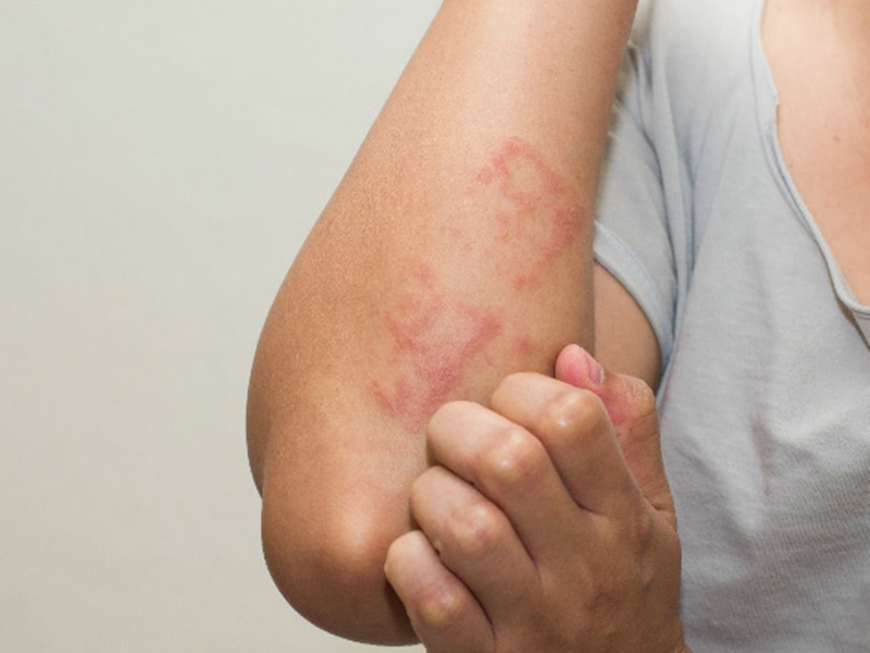
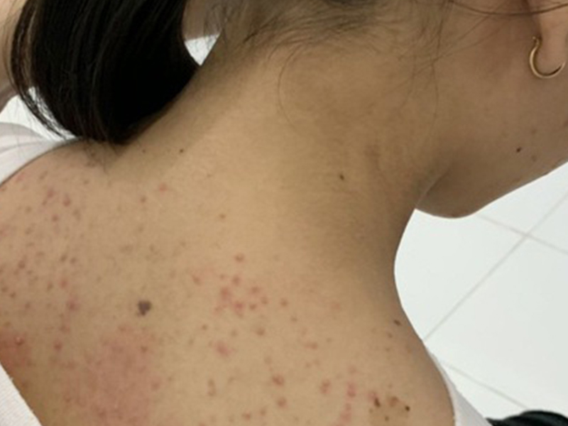
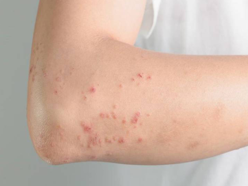
Common symptoms are myalgia, fever, bronchospasm, transient erythematous rash, lymphadenopathy, subcutaneous lumps with eosinophilia and, rarely, increased creatine kinase.
Symptomatic symptom-symptomatic symptomatic symptomatic symptomatic symptomatic symptomatic symptomatic symptomatic symptomatic symptomatic cardiopathy These follicles in the muscle have a very large size change, the diameter from under 50 - 325 of parasite in the heart muscle. The follicles in the muscles vary in size, with a diameter of less than 50 - 325 µm, the length of the follicles is up to 5cm.
Sarcocystis disease Intestinal sarcocystis
Patients presenting with gastrointestinal disorders, such as abdominal pain, diarrhea that resolves on their own, patients with gastrointestinal disorders, such as abdominal pain, diarrhea that resolves on their own, vomiting, fever, tremor, cysts or cells found Feces in stool.
5. DIAGNOSTIC REGISTRATION OF SARCOCYSTIS
- Stool test can see cyst spores * Stool test can see cyst spores in symptomatic and asymptomatic individuals.
- Patients with myalgia, muscle biopsy may see many follicles of Sarcocystis.
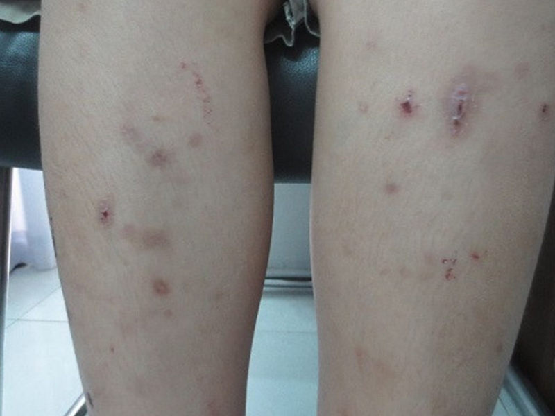
- Serum d. . Dịch vụ: Thiết kế website, quảng cáo google, đăng ký website bộ công thương uy tín
Related news
-
 Parasitical Worms.com Tests to find the cause of urticaria, diagnosis of urticaria results will be available throughout the day. After the results the doctor will explain, point out the abnormal signs for your child to understand and he will prescribe medication for home. Question Hello doctor: I ...
 Parasitical Worms.com Adult flukes are very small, 3 - 6 mm long, with 4 suction heads and a double hook, very short neck; coal consists of 3 segments, the final flukes have several hundred eggs, size 45 x 35 mcm, very similar to Toenia spp eggs. The disease is caused by the larva Echinococcus ...
 Parasitical Worms.com Some diseases caused by larvae of the anisakinae family parasitize marine mammals. In humans, the parasite falls into a dead-end, or severe or severe illness depending on the place of parasite, number of larvae and tissue responses. Diagnosis is often difficult and the most ...
 Parasitical Worms.com Illness caused by the nematode of Angiostrongylus cantonensis parasitizes and causes disease in the meninges, invasion of the brain can lead to death. Commonly called Meningitis - brain caused by Angiostrongylus cantonensis. The causative agent of nematode ...
 Fascioliasis is two types of fascioliasis and small liver fluke. People are infected with food, skin. Flukes can cause hepatitis, liver tumors, liver necrosis, but fortunately, liver fluke can be cured if detected early, treated in a reputable facility with a good doctor, using drugs. Good, ...
 Parasitical Worms.com Diagnosis is determined by seeing sparganum larvae from the wound. Clinical and prehistoric images of frog meat, eye-copying as well as the habit of eating undercooked snakes, mice, and eels are important factors for diagnosis. Doctor: Le Thi Huong Giang Medical Consultation: ...
 MUSHROOM DISEASE (Aspergillus) 1. Epidemiology. Aspergillus fungus is one of the largest fungal strains, present in all over the world, there are about 100 species, currently there are about 20-30 species that cause disease in humans, important strains are A. fumigatus, A. flavus , A. niger such as ...
 MUSHROOM DISEASE Cryptococcosis (Tolurosis, European Blastomycois) 1. Etiology and epidemiology Cryptococcosis is also known as the European Blastomycose mycosis caused by Cryptoccocus neoformans, a thick cystic yeast, has serotypes A, D (C. neoformans var. Neoformans) and B, C ( C.neoformans var. ...
 MUSHROOM DISEASE Sporotrichosis (Gardener Disease) 1. Epidemiology and etiology Sporotrichosis is a chronic disease caused by Sporothrix schenckii that causes damage to the skin or internal organs (also known as gardener disease - gardener's disease). This is a dimorphic mushroom. In nature, ...
 CANDIDA MUSHROOM 1. Germs Candidiasis is an acute, subacute or chronic disease caused by Candida-like yeasts, mostly Candida albicans. Candidiasis is available in the body (bronchus, oral cavity, intestine, vagina, skin around the anus) normally in non-pathogenic form. When having favorable ...

