Round Angiostrongylus Disease
Parasitical Worms.com Angiostrongylus cantonensis also known as vascular worms or synonyms Parastrongylus cantonensis, according to the parasitic worm parasitic taxonomy, this is a roundworm.
Worm infections caused by ingestion of undercooked, uncooked molluscs or freshwater snails are also the major intermediate hosts of this parasite or green vegetables contaminated with larvae in the mucus of snails, slugs or other flat creatures stick to the ground.
The larvae that migrate to the meninges appear to have meningeal or cortical symptoms. When the worms are almost adults, they move to the pulmonary artery branches
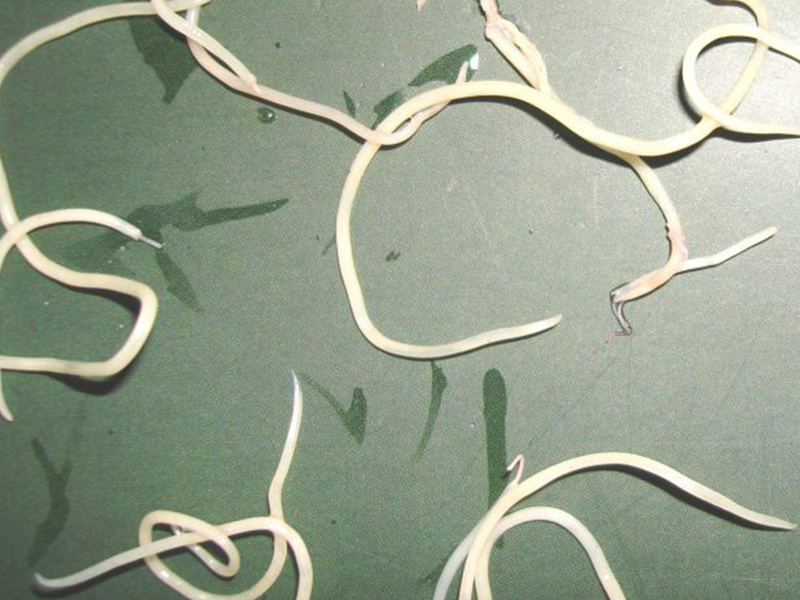
Images of the nematode worm Angiostrongylus
Distribution of angiostrongylus cantonensi vascular disease
According to an overall study by John H. Cross, of the Department of Health Sciences, Hebert University of Medicine, Bethesda, Maryland, the United States summarized the species of Angiostrongylus cantonensis, which is widely distributed in the Philippines, Indonesia, Malaysia, and Thailand
In the United States, this parasite has been found in rats, molluscs, and a reported case from New Orleans; In Africa (Madagascar) A.
 .cantonensis was detected in mice.
In Vietnam, since 1960, the number of suspected meningitis caused by A. cantonensis has been recorded and reported, especially in the case of autopsy patients detecting this worm (Phan Trinh and cs., 1974).
Since then, the number of detected cases has increased, approximately 70-100 cases are detected every year nationwide, the number of detected cases is too low compared to the number of cases tested positive for many times
Angiostrongylus cantonensis morphology morphology
A sample of adult worms A. Acantensis is located inside the pulmonary blood vessels. Through staining, the parasite A.
 .cantonensis the adult size is relatively, the male 20-22mm x 320-420µm, the female 22-34mm x 340-560µm, lives inside the capillary of the rat's lungs and there they lay eggs, then produce stage 1 larvae (L1).
The biological cycle of Angiostrongylus cantonensis
A. cantonensis parasites in some terrestrial snails (Achatina fulica, the large snail in Africa), aquatic snails (Pila spp., Viviparus javanica), or slugs (Veronicella alte and siamensis) can be closed role as an intermediate host for the development of L3 larvae. The existence of major intermediate hosts is described: in freshwater shrimp, land crabs and frogs that survive on snails and snails, the larvae remain infectious (L3) in some stages and may infecting mice, or humans when eating intermediate hosts such as Argentina fulica.
An infected person by ingesting uncooked snails, slugs, vegetables, or hosts; In humans, the larvae migrate to the central nervous system, where their growth stops and causes eosine-meningococcal meningitis.
When they lay and release the first stage larvae (L1), the larvae move to the small intestine through the alveolar, trachea, pharynx and esophageal cavities; then the larvae eliminate with faeces. The L1 larvae infect the first intermediate hosts, molluscs (snails and slugs), where they develop into the third-stage infectious larvae (L3).
Infected mice by ingesting snails or slugs with L3 larvae, the larvae in mice will move from the intestines to the central nervous system and mature there undergoing 2 developmental stages in 2 to 3 times.
 . week. Next, the adult worms move to the subarachnoid space, into the venous system, into the pulmonary artery and mature to the complete stage Larvae can be detected in mouse faeces within 40-60 days of infection.
Angiostrongylus cantonensis morphology morphology:
Cutting A.cantonensis worm in the subarachnoid cavity with inflammation and hemorrhage (when hematoxylin and eosin staining), A.cantonensis pathogenesis depends on the direct damage caused by the larvae that move and the young worm ( 7-13mm x 100-260µm long) and inflammatory granulomatous reaction.
Histological examination revealed slices of the worm surrounded by inflammatory cells (including neutrophils and eosinophils), capillary congestion, subarachnoid and hemorrhagic hemorrhage, localized necrosis and hemorrhage in the brain. The cross section of A. cantonensis adult worms in the brain has no inflammatory response; An incision of immature adult worms, A.Cantonensis lesions in the lungs or in the brain are also clearly seen through hematoxyli staining.
 . . Dịch vụ: Thiết kế website, quảng cáo google, đăng ký website bộ công thương uy tín
Related news
-
 Parasitical Worms.com Tests to find the cause of urticaria, diagnosis of urticaria results will be available throughout the day. After the results the doctor will explain, point out the abnormal signs for your child to understand and he will prescribe medication for home. Question Hello doctor: I ...
 Parasitical Worms.com Adult flukes are very small, 3 - 6 mm long, with 4 suction heads and a double hook, very short neck; coal consists of 3 segments, the final flukes have several hundred eggs, size 45 x 35 mcm, very similar to Toenia spp eggs. The disease is caused by the larva Echinococcus ...
 Parasitical Worms.com Some diseases caused by larvae of the anisakinae family parasitize marine mammals. In humans, the parasite falls into a dead-end, or severe or severe illness depending on the place of parasite, number of larvae and tissue responses. Diagnosis is often difficult and the most ...
 Parasitical Worms.com Illness caused by the nematode of Angiostrongylus cantonensis parasitizes and causes disease in the meninges, invasion of the brain can lead to death. Commonly called Meningitis - brain caused by Angiostrongylus cantonensis. The causative agent of nematode ...
 Fascioliasis is two types of fascioliasis and small liver fluke. People are infected with food, skin. Flukes can cause hepatitis, liver tumors, liver necrosis, but fortunately, liver fluke can be cured if detected early, treated in a reputable facility with a good doctor, using drugs. Good, ...
 Parasitical Worms.com Diagnosis is determined by seeing sparganum larvae from the wound. Clinical and prehistoric images of frog meat, eye-copying as well as the habit of eating undercooked snakes, mice, and eels are important factors for diagnosis. Doctor: Le Thi Huong Giang Medical Consultation: ...
 MUSHROOM DISEASE (Aspergillus) 1. Epidemiology. Aspergillus fungus is one of the largest fungal strains, present in all over the world, there are about 100 species, currently there are about 20-30 species that cause disease in humans, important strains are A. fumigatus, A. flavus , A. niger such as ...
 MUSHROOM DISEASE Cryptococcosis (Tolurosis, European Blastomycois) 1. Etiology and epidemiology Cryptococcosis is also known as the European Blastomycose mycosis caused by Cryptoccocus neoformans, a thick cystic yeast, has serotypes A, D (C. neoformans var. Neoformans) and B, C ( C.neoformans var. ...
 MUSHROOM DISEASE Sporotrichosis (Gardener Disease) 1. Epidemiology and etiology Sporotrichosis is a chronic disease caused by Sporothrix schenckii that causes damage to the skin or internal organs (also known as gardener disease - gardener's disease). This is a dimorphic mushroom. In nature, ...
 CANDIDA MUSHROOM 1. Germs Candidiasis is an acute, subacute or chronic disease caused by Candida-like yeasts, mostly Candida albicans. Candidiasis is available in the body (bronchus, oral cavity, intestine, vagina, skin around the anus) normally in non-pathogenic form. When having favorable ...


