Small Liver Fluke- Large Liver Fluke
Small liver fluke: there will be 3 types: Clonorchis sinensis, Opisthorchis viverrini; Opisthorchis felineus
Flukes of the liver: there are two types: Fasciola hepatica and Fasciola gigantica
The flukes are in the liver
Shape and size: Fascioliasis and small liver fluke are both shaped like leaves, thin banks and flat bodies. Size varies depending on the type: large liver fluke has a larger size than small liver fluke. The body of the flukes is bisexual, it has both ovaries and testes on a fluke.
Probability of survival in the external environment: Eggs of liver fluke have a thin shell, so the external environment is very low, at a temperature of sunlight above 70 ° C the eggs will be damaged. However, if the liver fluke eggs want to develop into larvae, they must be in water because the eggs will be damaged and cannot grow on land, the possibility of small liver fluke maturation short
2. What will a person infected with liver fluke look like?
Small liver fluke disease:
The main cause is humans and in some animals such as cats, dogs, tigers, foxes, otters, rats, insects on the ground in addition to the first vectors of transmission of snails and hosts the second vector are freshwater fish .
People infected by eating fish and snails containing uncooked tapeworm larvae should eat them into the stomach, duodenum and then back up the bile to the liver, thus developing parasites. in the liver and cause biliary tract disease.
 .
.Adult life cycle of a giant liver fluke
Fascioliasis
The cause is mainly herbivores such as buffaloes, cows, vectors that are transmitted by snails, which people often eat.
People who are infected by eating raw vegetables (cilantro, parsley, celery, mustard greens) or drinking rain or water may not be clean with tapeworm larvae.
3. Symptoms of liver fluke symptoms like?
3.1
The incubation period for small fluke and large liver fluke will depend on the number of larvae encountered by people, for small liver fluke infection with more than 100 new flukes, it is possible to show markedly, for fluke Large liver incubation period is difficult to determine accurately and there is no outward manifestation.
3.2. Period of transmission
For small liver fluke: After the parasite is penetrated into the liver parenchyma, the young tapeworm will develop into an adult fluke and be laid in bile ducts, the eggs are excreted in the feces into the water and distributed.
 . The growth in a cycle thus spreads through live cystic fish.
Because liver fluke is parasitic in the bile ducts, when infected with small liver fluke may have symptoms: Symptoms of regional pain in the liver due to reproductive fluids cause obstruction of the bile ducts in the liver thus leading to the manifestations Pain in the lower right flank.
Anorexia, flatulence, digestive disorders, nausea.
There are also signs of dullness, jaundice, yellow eyes and signs of enlarged liver or cirrhosis, depending on the severity of the disease
In the worst case, small flukes can occur, which can cause biliary tract inflammation, biliary tract hemorrhage, biliary tract cancer, and biliary cirrhosis.
For large liver flukes:
After 2 to 3 months of infiltration into the liver parenchyma, the tapeworm will move into the bile ducts, mature and form eggs, the eggs will go out in the fecal route followed by the water. The larvae pass through the snails and develop into larvae and cysts that attach to the vegetables or swim in the water, so if a person or herbivore is eaten, the larvae will enter the stomach and enter the intestine, and then enter the liver and sign. born at the liver. At mature liver fluke can parasitize and cause disease in people for many years.
When infected with large liver fluke patients often have one or the following manifestations:
Pain in the lower flank must be spread backward or epigastric, manifesting nonspecific pain, may be shady with intermittent episodes, sometimes severe pain, there will be cases without abdominal pain.
 .
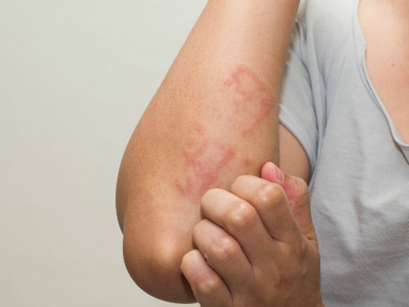
.Patients who are often tired, have dyspepsia, dyspepsia, nausea, may have a slight fever or aching joints, muscle aches, and urticaria.
In addition, some cases of massive liver fluke can cause liver abscess, patients with severe right flank pain, high fever, and enlarged liver If the abscess bursts into the lungs, it can cause pleural effusion, severe and dangerous port disease.
Some cases of parasitic parasites will be stray like the lungs, the skin under the chest ....
Symptoms of liver fluke disease are similar to other liver diseases such as viral hepatitis, cholangitis, liver cancer, or liver abscess due to other causes. It is necessary to carefully examine and test for appropriate treatment.
4.
 How to know if you have liver fluke infection?
If you suspect you have infection with liver fluke, you can go to a hospital or clinic for testing:
Small liver fluke disease: a test found in a stool or duodenum egg.
Fascioliasis: a test found in faecal eggs or blood tests by immunological techniques (ELISA) can find antibodies to large liver fluke in the serum of patients. . Dịch vụ: Thiết kế website, quảng cáo google, đăng ký website bộ công thương uy tín
Related news
-
 Parasitical Worms.com Tests to find the cause of urticaria, diagnosis of urticaria results will be available throughout the day. After the results the doctor will explain, point out the abnormal signs for your child to understand and he will prescribe medication for home. Question Hello doctor: I ...Parasitical Worms.com Adult flukes are very small, 3 - 6 mm long, with 4 suction heads and a double hook, very short neck; coal consists of 3 segments, the final flukes have several hundred eggs, size 45 x 35 mcm, very similar to Toenia spp eggs. The disease is caused by the larva Echinococcus ...Parasitical Worms.com Some diseases caused by larvae of the anisakinae family parasitize marine mammals. In humans, the parasite falls into a dead-end, or severe or severe illness depending on the place of parasite, number of larvae and tissue responses. Diagnosis is often difficult and the most ...
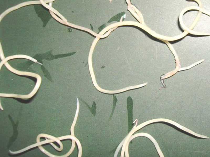 Parasitical Worms.com Illness caused by the nematode of Angiostrongylus cantonensis parasitizes and causes disease in the meninges, invasion of the brain can lead to death. Commonly called Meningitis - brain caused by Angiostrongylus cantonensis. The causative agent of nematode ...Fascioliasis is two types of fascioliasis and small liver fluke. People are infected with food, skin. Flukes can cause hepatitis, liver tumors, liver necrosis, but fortunately, liver fluke can be cured if detected early, treated in a reputable facility with a good doctor, using drugs. Good, ...
 Parasitical Worms.com Diagnosis is determined by seeing sparganum larvae from the wound. Clinical and prehistoric images of frog meat, eye-copying as well as the habit of eating undercooked snakes, mice, and eels are important factors for diagnosis. Doctor: Le Thi Huong Giang Medical Consultation: ...
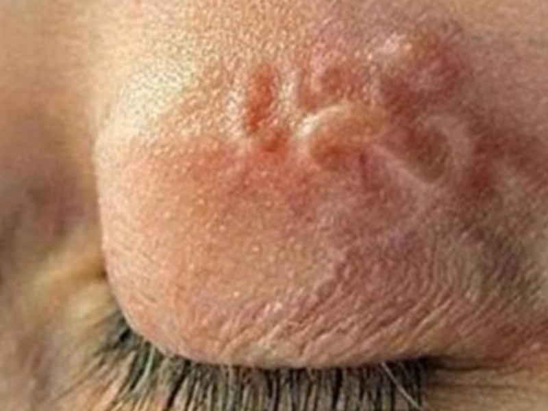 MUSHROOM DISEASE (Aspergillus) 1. Epidemiology. Aspergillus fungus is one of the largest fungal strains, present in all over the world, there are about 100 species, currently there are about 20-30 species that cause disease in humans, important strains are A. fumigatus, A. flavus , A. niger such as ...
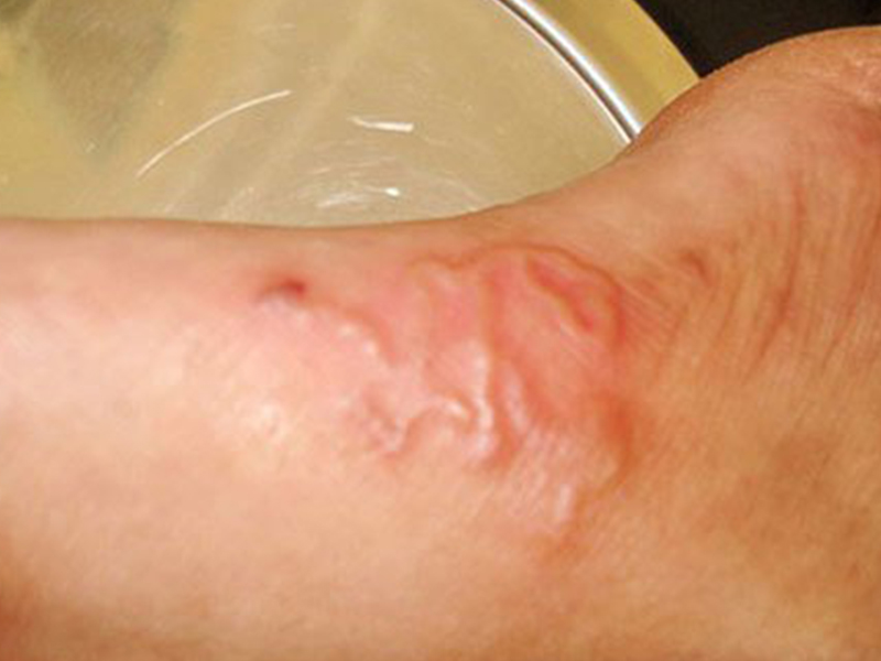 MUSHROOM DISEASE Cryptococcosis (Tolurosis, European Blastomycois) 1. Etiology and epidemiology Cryptococcosis is also known as the European Blastomycose mycosis caused by Cryptoccocus neoformans, a thick cystic yeast, has serotypes A, D (C. neoformans var. Neoformans) and B, C ( C.neoformans var. ...
 MUSHROOM DISEASE Sporotrichosis (Gardener Disease) 1. Epidemiology and etiology Sporotrichosis is a chronic disease caused by Sporothrix schenckii that causes damage to the skin or internal organs (also known as gardener disease - gardener's disease). This is a dimorphic mushroom. In nature, ...CANDIDA MUSHROOM 1. Germs Candidiasis is an acute, subacute or chronic disease caused by Candida-like yeasts, mostly Candida albicans. Candidiasis is available in the body (bronchus, oral cavity, intestine, vagina, skin around the anus) normally in non-pathogenic form. When having favorable ...