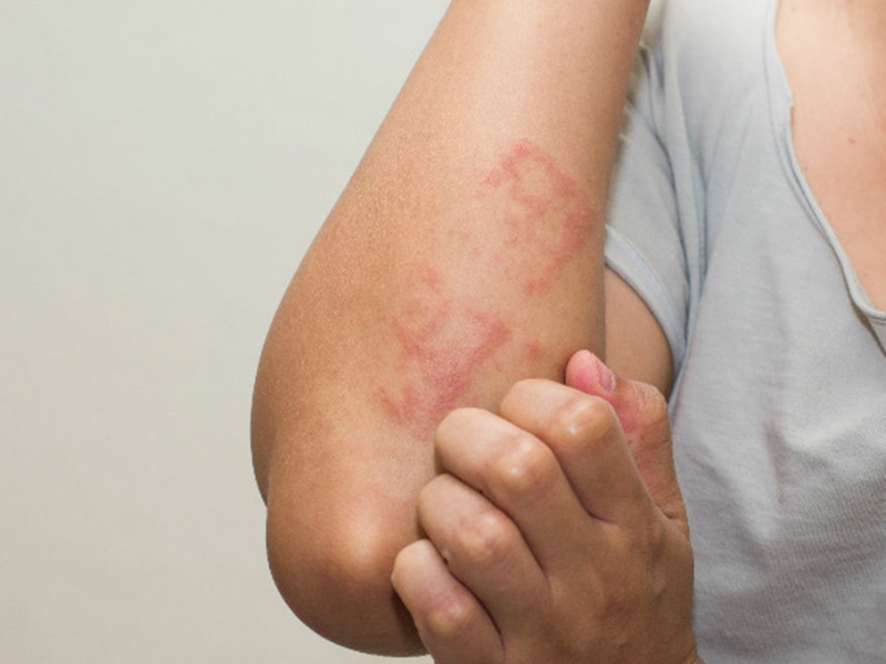
Strongyloides Strongyloidiasis Identification Sign
Parasitical Worms.com Strongyloidiasis was discovered by Louis Normand in 1876 causing digestive disorders, especially diarrhea. The first cases were encountered in French soldiers in Indochina. After many name changes, from 1911 until now the pathogen has been uniformly named Strongyloides stercoralis.
There are 52 different species of strongyloidiasis, but only two species that cause disease in humans are Strongyloides stercoralis and Strongyloides fuelleborni
1 Strongyloides Strongyloid form
Strongyloides parasitic stercoralis
Female worms are tubular, very small, length 2 -2.8mm, horizontally about 37 - 40 µm, transparent.
 . The mouth has two lips, the esophagus is about 1/4 of the length of the whole body. The genitourinary system of Strongyloides stercoralis consists of the uterus and ovaries lying symmetrically through the genital opening in the middle of the worm body. Female worms live in the layer under the mucous membrane of the human digestive tract.
No parasitic male worms have been found. Some hypotheses that there are still parasitic males but very small can not be found
Strongyloides stercoralis lives freely
Female worms are about µ - 1.5mm long, 50-80 µm wide, and the esophagus is bulging.
Free-living male worms are shorter than females, J-shaped, about 0.
 .7mm long, about 50 ngangm across, curved tails with two genital spines.
Strongyloides stercoralis eel eggs
Strongyloides stercoralis egg (parasitic) oval, about 54 x 32 µm, thin, transparent shell, like hookworm eggs but with larvae available
The eggs laid by females that live freely will have a larger size, 70 x 45 µm, the shell is just a thin film, can change shape from round to oval as the larvae move inside the egg.
Image of two types of strongyloid larvae
Larvae stage I (rhabditiform - larvae with distended esophagus): Hatched from eggs, about 200 - 250 x 16 - 20 µm, short mouth sinuses, pointed tail, esophagus with tight waist, should be bulge.
Stage 2 larvae (filariform - larvae with tubular esophagus): Developed from stage I Larvae vary in size from 400 to 700 µm, across 12-20 µm, and the esophagus has a long tubular shape from 40% - 45% of body length. Tail tail or forked at the end like a swallow.
2. Strongyloides strongyloidiasis development cycle
People who are exposed to faecal-contaminated soil have their larvae
Stage 2 (filariform) live freely from the skin. The larvae travel through the bloodstream and travel to the lungs through the circulation. In the lungs, the parasite breaks down the pulmonary capillaries and enters the alveoli, then they move to the bronchi, trachea, into the pharynx, esophagus and are swallowed into the digestive tract, developing into adult female worms.
 . .
. .Adult female worms live parasitically, parasitizing the intestinal mucosa, laying eggs Eggs hatch into stage I larvae in the intestinal mucosa, excreting in the stool. Stage 1 larvae develop into stage 2 larvae, penetrating the skin upon contact with the soil, similar to hookworms (direct cycle).
Larvae 2 can switch to an indirect cycle (free life), molting four times to males and adult females living freely. Adults mate and create generations of descendants to the next parasitic life similar to the direct cycle.
Favorable conditions within the host and disadvantages outside the host easily for the direct cycle; unfavorable conditions inside the host and favorable outside the host easily for the next cycle.
A feature of Strongyloides stercoralis is that a small part of stage 1 larvae molts in the small intestine into stage 2 larvae. Stage 2 larvae penetrate the wall of the large intestine or perianal skin, completing the lateral cycle. in the human body, developing into males and females maturing in the small intestine.

This is a self-infecting cycle, this cycle occurs regularly and continuously, making the host body always have circulating larvae, lasting for months, years, although the host is not re-infected. This autoimmune cycle is responsible for many complex medical problems. This cycle predominates in immunocompromised individuals such as corticoids abuse, HIV infection, HTLV-1, leukemia, organ transplantation.
In addition to the normal way, they can follow the bloodstream to penetrate various organs in the body. Depending on the parasite position of the larvae in the body, patients will have different clinical manifestations in their respective organs. In these cases, the larvae are rarely detected in the stool, so the diagnosis must be based on immunological serology.
3. Epidemiological characteristics of Strongyloides strongyloidiasis
Sturchler in 1981 divided the situation of strongyloidiasis into three areas: mild endemic area when the prevalence is <1%, endemic area from 1% - 5% and severe endemic area> 5%. . Dịch vụ: Thiết kế website, quảng cáo google, đăng ký website bộ công thương uy tín
Related news
-
 Parasitical Worms.com Tests to find the cause of urticaria, diagnosis of urticaria results will be available throughout the day. After the results the doctor will explain, point out the abnormal signs for your child to understand and he will prescribe medication for home. Question Hello doctor: I ...Parasitical Worms.com Adult flukes are very small, 3 - 6 mm long, with 4 suction heads and a double hook, very short neck; coal consists of 3 segments, the final flukes have several hundred eggs, size 45 x 35 mcm, very similar to Toenia spp eggs. The disease is caused by the larva Echinococcus ...
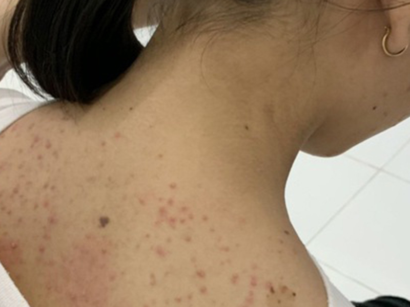 Parasitical Worms.com Some diseases caused by larvae of the anisakinae family parasitize marine mammals. In humans, the parasite falls into a dead-end, or severe or severe illness depending on the place of parasite, number of larvae and tissue responses. Diagnosis is often difficult and the most ...
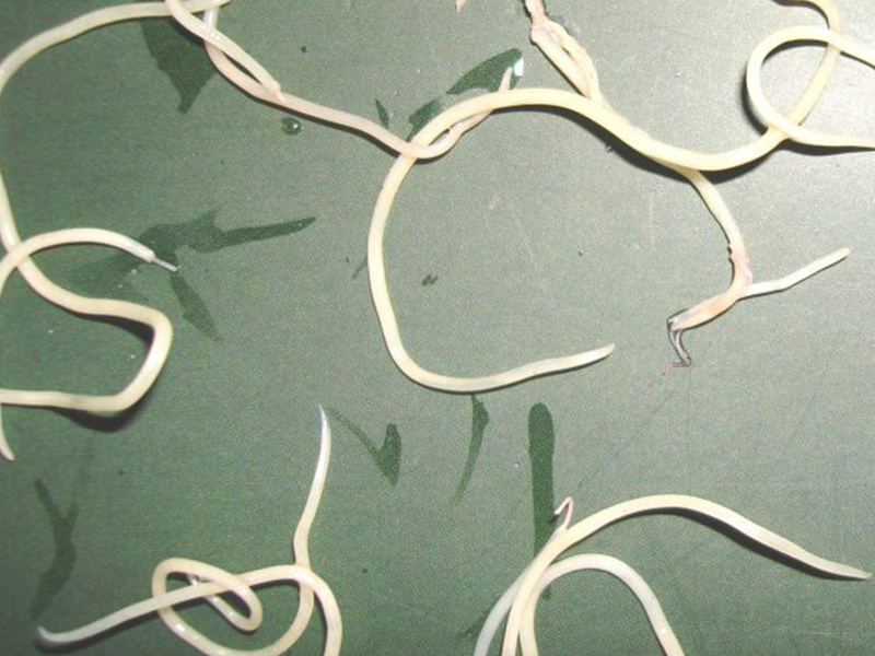 Parasitical Worms.com Illness caused by the nematode of Angiostrongylus cantonensis parasitizes and causes disease in the meninges, invasion of the brain can lead to death. Commonly called Meningitis - brain caused by Angiostrongylus cantonensis. The causative agent of nematode ...Fascioliasis is two types of fascioliasis and small liver fluke. People are infected with food, skin. Flukes can cause hepatitis, liver tumors, liver necrosis, but fortunately, liver fluke can be cured if detected early, treated in a reputable facility with a good doctor, using drugs. Good, ...Parasitical Worms.com Diagnosis is determined by seeing sparganum larvae from the wound. Clinical and prehistoric images of frog meat, eye-copying as well as the habit of eating undercooked snakes, mice, and eels are important factors for diagnosis. Doctor: Le Thi Huong Giang Medical Consultation: ...
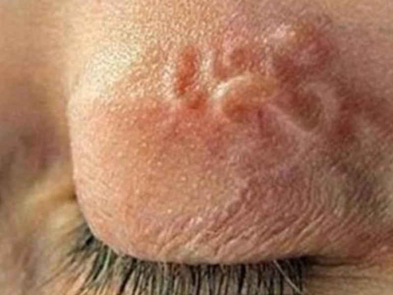 MUSHROOM DISEASE (Aspergillus) 1. Epidemiology. Aspergillus fungus is one of the largest fungal strains, present in all over the world, there are about 100 species, currently there are about 20-30 species that cause disease in humans, important strains are A. fumigatus, A. flavus , A. niger such as ...
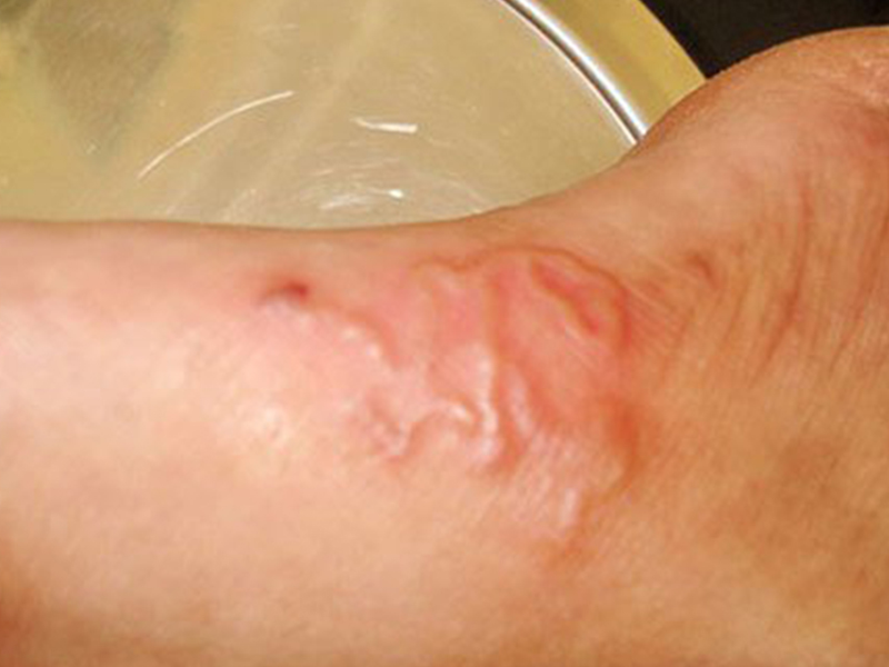 MUSHROOM DISEASE Cryptococcosis (Tolurosis, European Blastomycois) 1. Etiology and epidemiology Cryptococcosis is also known as the European Blastomycose mycosis caused by Cryptoccocus neoformans, a thick cystic yeast, has serotypes A, D (C. neoformans var. Neoformans) and B, C ( C.neoformans var. ...MUSHROOM DISEASE Sporotrichosis (Gardener Disease) 1. Epidemiology and etiology Sporotrichosis is a chronic disease caused by Sporothrix schenckii that causes damage to the skin or internal organs (also known as gardener disease - gardener's disease). This is a dimorphic mushroom. In nature, ...CANDIDA MUSHROOM 1. Germs Candidiasis is an acute, subacute or chronic disease caused by Candida-like yeasts, mostly Candida albicans. Candidiasis is available in the body (bronchus, oral cavity, intestine, vagina, skin around the anus) normally in non-pathogenic form. When having favorable ...