Tapeworm Disease
Parasitical Worms.com When humans or other animals eat or swallow tapeworm eggs, the larvae are released into the duodenum and enter the intestinal wall, along veins, veins, and into the circulatory system throughout the body. .
I. Physical characteristics of dog flukes
Dog flukes (tapeworms) have the scientific name of Echinococcus granulosus about 3 to 6 mm long, pear-shaped head, about 0.3 mm horizontally. The body consists of 3 to 4 segments
 .
.Adult dog tapeworms and tapeworm eggs
The main hosts of tapeworms are domestic dogs, jackals, sometimes foxes. After the parasite develops in the dog's intestines, the old flukes are burned automatically to the outside of the anus and broken causing the tapeworm eggs to spread everywhere. When burning flukes out of the anus stimulates itching, the dog licks the anus and licks the fur so the dog's fur also gets lots of tapeworm eggs and easily infects other hosts such as sheep, buffaloes, cows, horses, goats , pig. The sheep is the main secondary host and the person is the random secondary host.
The life cycle of a dog flukes
When humans or other animals eat or swallow tapeworm eggs, larvae are released into the duodenum and enter the intestinal wall, along veins, veins, into the circulatory system throughout the body
Cystic tapeworm and cystic tapeworm
There are 3 types of cysts in humans including aunilocular cysts (aunilocular), bone cysts (osseous) that develop in bone tissue and cysts (alveolar) of Echinococcus multilocularis. This type of single-follicular capsule is common in humans, less common in animals.
 . Cysts develop slowly for many years, round; common with 66% in liver, 22% in lungs; 3% in kidney, 2% in bone, 1% in brain and some other organs such as muscles, spleen, heart, eyes.
Cystic tapeworm in the liver
The structure of a hydatidcyst consists of a crust of about 1 mm thick and a reproductive film from 22 to 25 dàym thick, inside which is a yellowish fluid. The brood capsule has only the inner reproductive membrane containing flukes. Cystic tapeworm has a repeated structure of the mother fluke cyst
Cysticercosis in the brain
When the tapeworm explodes, many small flukes from the cysts drain out into the cyst fluid. An average tapeworm follicle contains about 2 million premature tapeworms. If the dog ate a tapeworm, after 7 weeks in the dog's body there are millions of adult flukes. If the tapeworm bursts in the host's body, the young tapeworm grows into a new cystic worm called a secondary tapeworm. Cysts in a tapeworm fluid can sometimes produce a tapeworm cyst.
Some cysts are calcified or invaded by bacteria, they do not have hatching cysts and do not have flukes called "clean" or headless cysts (acephalocysts).
 .
.Pathological symptoms, diagnosis and treatment of disease
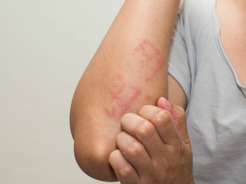
When Echinococcus granulosus invades and parasites in the body, the tapeworm will compress the surrounding organs and organs, causing serious complications. The damage and harm also depend on the location of the parasitic cyst
If the tapeworm is broken, it causes intoxication, allergies, anaphylaxis, and the flukes spill out and form secondary cysts. Secondary cysts may appear 2 to 5 years after the first appearance of the primary cysts is broken and often fatal at this stage.
The diagnosis of a tapeworm cyst is often difficult because the cysts grow slowly compared to other types of cysts. Therefore, in clinical practice, the tapeworm usually does not detect in time as the case of the tapeworm in the palate may take up to 30 years to show severe symptoms. Through X-ray techniques, it is possible to detect early cysts. Blood tests that show eosinophilia to increase by 20 to 25% or a fluke-specific immune serological diagnosis that give positive results are important indicators and directions for the diagnosis.
Images of supercooled dog flukes and on XQ film
Treating schistosomiasis by surgical method is applied to those cysts which can be operated and peeled whole. In the case of inoperable cysts, biological therapy is given by repeatedly injecting the patient with fluid removed from hydatid cysts, presumably injecting an antigen; gradually the cyst flukes in the patient will be scaled down.
People suffering from schistosomiasis need to be monitored and treated at a medical facility with a parasite specialty
Preventing tapeworm disease
Although Echinococcus granulosus is rarely detected in our country, according to the literature and Faust Scientist, 1970 mentioned, the study noted this distribution, present in some Asian countries like China, Japan, the Philippines and Vietnam. So scientists and the health care industry should care and do not forget a rare parasitic disease but it is very likely to be transmitted from domestic dogs. . Dịch vụ: Thiết kế website, quảng cáo google, đăng ký website bộ công thương uy tín
Related news
-
 Parasitical Worms.com Tests to find the cause of urticaria, diagnosis of urticaria results will be available throughout the day. After the results the doctor will explain, point out the abnormal signs for your child to understand and he will prescribe medication for home. Question Hello doctor: I ...Parasitical Worms.com Adult flukes are very small, 3 - 6 mm long, with 4 suction heads and a double hook, very short neck; coal consists of 3 segments, the final flukes have several hundred eggs, size 45 x 35 mcm, very similar to Toenia spp eggs. The disease is caused by the larva Echinococcus ...
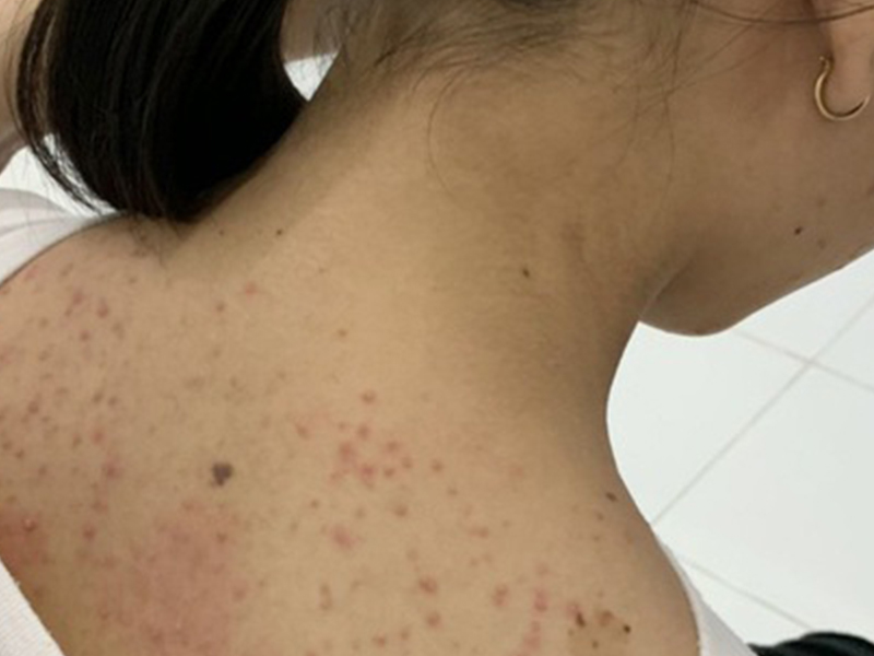 Parasitical Worms.com Some diseases caused by larvae of the anisakinae family parasitize marine mammals. In humans, the parasite falls into a dead-end, or severe or severe illness depending on the place of parasite, number of larvae and tissue responses. Diagnosis is often difficult and the most ...
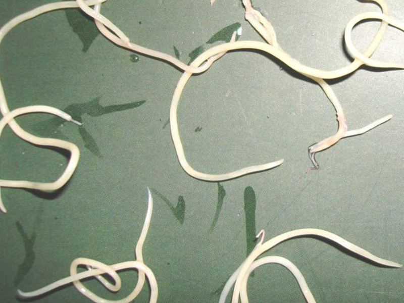 Parasitical Worms.com Illness caused by the nematode of Angiostrongylus cantonensis parasitizes and causes disease in the meninges, invasion of the brain can lead to death. Commonly called Meningitis - brain caused by Angiostrongylus cantonensis. The causative agent of nematode ...Fascioliasis is two types of fascioliasis and small liver fluke. People are infected with food, skin. Flukes can cause hepatitis, liver tumors, liver necrosis, but fortunately, liver fluke can be cured if detected early, treated in a reputable facility with a good doctor, using drugs. Good, ...
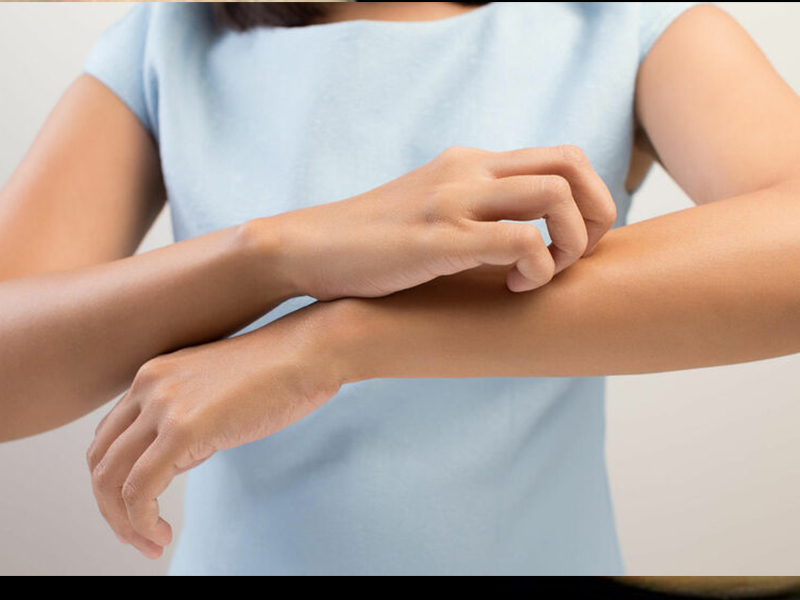 Parasitical Worms.com Diagnosis is determined by seeing sparganum larvae from the wound. Clinical and prehistoric images of frog meat, eye-copying as well as the habit of eating undercooked snakes, mice, and eels are important factors for diagnosis. Doctor: Le Thi Huong Giang Medical Consultation: ...
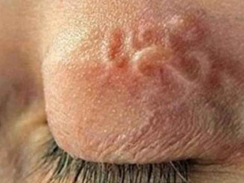 MUSHROOM DISEASE (Aspergillus) 1. Epidemiology. Aspergillus fungus is one of the largest fungal strains, present in all over the world, there are about 100 species, currently there are about 20-30 species that cause disease in humans, important strains are A. fumigatus, A. flavus , A. niger such as ...
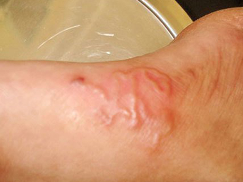 MUSHROOM DISEASE Cryptococcosis (Tolurosis, European Blastomycois) 1. Etiology and epidemiology Cryptococcosis is also known as the European Blastomycose mycosis caused by Cryptoccocus neoformans, a thick cystic yeast, has serotypes A, D (C. neoformans var. Neoformans) and B, C ( C.neoformans var. ...
 MUSHROOM DISEASE Sporotrichosis (Gardener Disease) 1. Epidemiology and etiology Sporotrichosis is a chronic disease caused by Sporothrix schenckii that causes damage to the skin or internal organs (also known as gardener disease - gardener's disease). This is a dimorphic mushroom. In nature, ...CANDIDA MUSHROOM 1. Germs Candidiasis is an acute, subacute or chronic disease caused by Candida-like yeasts, mostly Candida albicans. Candidiasis is available in the body (bronchus, oral cavity, intestine, vagina, skin around the anus) normally in non-pathogenic form. When having favorable ...