Where Do You Have To Treat?
Parasitical Worms.com Diagnostic tests to identify the environment of ringworm infection need to be done when treating patients. This is important because:) the duration of treatment; ii) high treatment costs; iii) Side effects of the drug after prolonged use. Therefore, treatment in the absence of disease will bring patients more costly and annoying.
The observation of mycelia and spores on wet specimens is sufficient to diagnose dermatophyte fungal infections but does not allow the identification of the causative agent except in case of fungal hair
Get the specimen
Of the specimens collected from suspected lesions caused by ringworm infection, about 50% of the samples were not found Therefore, it is necessary to ensure proper principles and techniques of specimen collection so that the test results can be accurate.
Rule
Patients should not take antifungal drugs within 7 - 10 days before taking samples for testing.
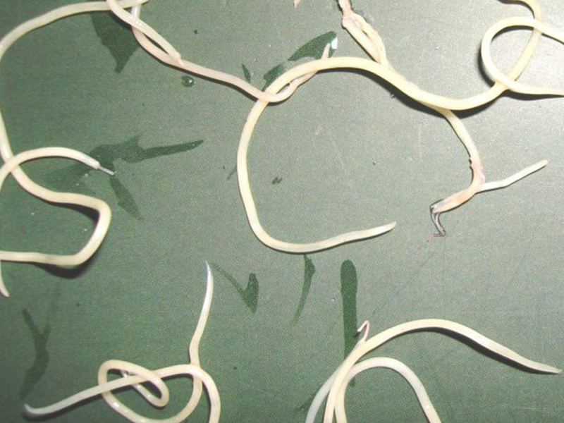 .
.Collect specimens from many different lesions and in different locations on the same lesion because the fungus is unevenly distributed on the lesions.
Fungus image
Collecting technique
Disinfect the affected area with 70% alcohol before removing the specimen to remove the debris, germs and pharmaceutical chemicals applied by the patient.
Collected specimens must be stored in dry condition to avoid the growth of germs and mold.
The type of specimen will vary depending on the clinical form.
Hair, cheek fungus: Pluck hairs of the beard, hairs are bent, lost shine, broken or fluorescent under the Wood light
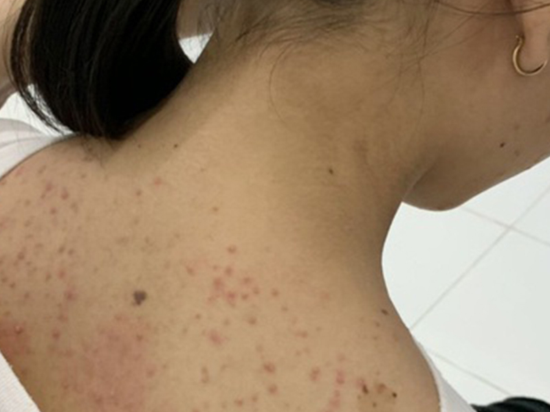
Fungal dermatitis: High skin scabs on the outside of the lesion with blue slides or scalpels are no longer sharp.
Onychomycosis: Specimens should be collected on discolored or dystrophy nails. For the distant fungal nail form, cut off the far edge of the nail and take in as much as possible, at the same time collecting additional layers of underside horn under the nail. In case of near-shore fungus, use a small drill or scalpel to remove the underlying nail.
 . Nail powder is collected on the nail surface in the form of white fungus.
Direct observation
Wet the specimen with 10% or 20% KOH solution and observe under a microscope to detect fungus.
Skin or nail specimen
Observations with objectives x 10 or x 40 will show on the skin epidermal cells or nails appear slender, branched, fungal hyphae and multiple spores. The test may be repeated several times when negative or clinically free of fungal pathogens
Specimens are beard, hair
Hair follicles get parasitic fungi in the following five types depending on the causative agent
Internal play classic style.
Internal concave style.
Broadcast - internal style microsporique
External distribution - internal trichophytique.
In the diagnosis of skin-borne fungal diseases, implantation is a more reliable method than direct observation because it allows the identification of pathogenic microorganisms, thus providing information about the source of the infection and knowledge of the source. will greatly assist in treatment.
Dermatological fungi have good resistance to some common antibacterial antibiotics such as chloramphenicol, gentamycine, penicillin, streptomycin .
 .. and with antifungal antibiotic cycloheximid (actidione), so when culturing specimens to isolate skin fungi, These antibiotics need to be mixed in culture to inhibit the growth of bacteria and fungal infection from the external environment.
However, it should be noted that some molds (Scytalidium, Scopulariopsis, Aspergillus, Aspergillus, Acremonium, Fusarium, etc.) may be the agents of nail inflammation and clinically difficult to distinguish from skin fungus infections. Therefore, it is advisable to inform the laboratory of suspected mold infections for inoculation on two media with and without cycloheximide.
The isolation of fungal pathogens started with Sabouraud chloramphenicol cycloheximide media incubated at room temperature.
Each specimen should be inoculated into two medium tubes. After 2–3 weeks, sometimes 1–1.5 months, the fungus develops into a cyst.
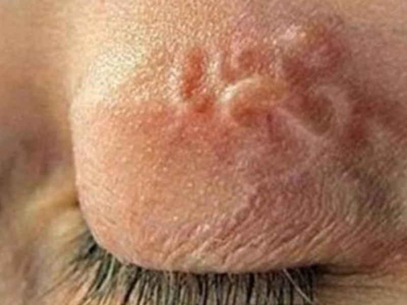 General observations of fungi and fungi will allow identification of fungi. In some cases, additional biological tests may be required to determine it more accurately
Inoculation of rice medium to distinguish M.canis from M.audouinii.
Use hair penetrationtest, urea, sweet potato agar medium to distinguish T.mentagrophytes and T.rubrum.
Inoculate Trichophyton medium 1 - 7 to identify Tconcentricum, T.tonsurans.
 .
.Distinguish the superficial varieties of fungi.
Dermatobacteria is a filamentous mycelium made of thin, baffled hyphae. In addition, some species with special structures such as racket-shaped mycelium, comb-shaped, deer-shaped mycelia, etc. are important characteristics for identification. Most of them produce attached spores (microconidi. . Dịch vụ: Thiết kế website, quảng cáo google, đăng ký website bộ công thương uy tín
Related news
-
 Parasitical Worms.com Tests to find the cause of urticaria, diagnosis of urticaria results will be available throughout the day. After the results the doctor will explain, point out the abnormal signs for your child to understand and he will prescribe medication for home. Question Hello doctor: I ...
 Parasitical Worms.com Adult flukes are very small, 3 - 6 mm long, with 4 suction heads and a double hook, very short neck; coal consists of 3 segments, the final flukes have several hundred eggs, size 45 x 35 mcm, very similar to Toenia spp eggs. The disease is caused by the larva Echinococcus ...
 Parasitical Worms.com Some diseases caused by larvae of the anisakinae family parasitize marine mammals. In humans, the parasite falls into a dead-end, or severe or severe illness depending on the place of parasite, number of larvae and tissue responses. Diagnosis is often difficult and the most ...
 Parasitical Worms.com Illness caused by the nematode of Angiostrongylus cantonensis parasitizes and causes disease in the meninges, invasion of the brain can lead to death. Commonly called Meningitis - brain caused by Angiostrongylus cantonensis. The causative agent of nematode ...
 Fascioliasis is two types of fascioliasis and small liver fluke. People are infected with food, skin. Flukes can cause hepatitis, liver tumors, liver necrosis, but fortunately, liver fluke can be cured if detected early, treated in a reputable facility with a good doctor, using drugs. Good, ...
 Parasitical Worms.com Diagnosis is determined by seeing sparganum larvae from the wound. Clinical and prehistoric images of frog meat, eye-copying as well as the habit of eating undercooked snakes, mice, and eels are important factors for diagnosis. Doctor: Le Thi Huong Giang Medical Consultation: ...
 MUSHROOM DISEASE (Aspergillus) 1. Epidemiology. Aspergillus fungus is one of the largest fungal strains, present in all over the world, there are about 100 species, currently there are about 20-30 species that cause disease in humans, important strains are A. fumigatus, A. flavus , A. niger such as ...
 MUSHROOM DISEASE Cryptococcosis (Tolurosis, European Blastomycois) 1. Etiology and epidemiology Cryptococcosis is also known as the European Blastomycose mycosis caused by Cryptoccocus neoformans, a thick cystic yeast, has serotypes A, D (C. neoformans var. Neoformans) and B, C ( C.neoformans var. ...
 MUSHROOM DISEASE Sporotrichosis (Gardener Disease) 1. Epidemiology and etiology Sporotrichosis is a chronic disease caused by Sporothrix schenckii that causes damage to the skin or internal organs (also known as gardener disease - gardener's disease). This is a dimorphic mushroom. In nature, ...
 CANDIDA MUSHROOM 1. Germs Candidiasis is an acute, subacute or chronic disease caused by Candida-like yeasts, mostly Candida albicans. Candidiasis is available in the body (bronchus, oral cavity, intestine, vagina, skin around the anus) normally in non-pathogenic form. When having favorable ...

